

doi: 10.1093/jmcb/mjz096.
Modulation of fatty acid synthase by ATR checkpoint kinase Rad3
Affiliations
- PMID: 31509190
- PMCID: PMC6934155
- DOI: 10.1093/jmcb/mjz096
Item in Clipboard
Modulation of fatty acid synthase by ATR checkpoint kinase Rad3
J Mol Cell Biol.
.
No abstract available
Figures
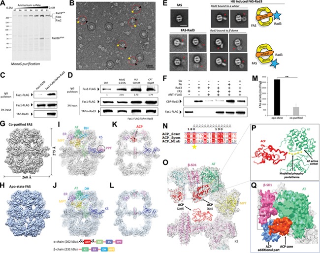
The interaction between S. pombe FAS and Rad3/Rad26 and structure/activity comparisons between co-purified and apo-state FAS. (A) Ruby-stained SDS–PAGE gel of Mono S chromatography fractions. (B) Negative stain EM micrograph of co-purified FAS–Rad3/Rad26. Black circles indicate FAS. Red circles indicate FAS–Rad3/Rad26 complex. Yellow and black arrows indicate Rad3/Rad26 and FAS, respectively in the complex. Scale bar, 50 nm. (C) In vivo co-IP assays in the Fas1-FLAG/TAPm-Rad3 double tagging strain and the Fas1-FLAG single tagging strain (negative control). TAPm-Rad3 has affinity with IgG resin and Fas1-FLAG was co-precipitated by TAPm-Rad3. Western blot analysis using indicated antibodies detecting FAS and Rad3. (D) Western blot of in vivo co-IP assays in cells treated with MMS, HU, and CPT. (E) 2D averages of apo-state FAS (left top panel), non-induced co-purified FAS–Rad3/Rad26 (left bottom panel) and HU-induced co-purified FAS–Rad3/Rad26 (right panel). Red arrows indicate Rad3/Rad26 density. Diagrams representing the typical complex configurations are shown beside the averages. Scale bar, 30 nm. (F) In vitro pull-down assays with affinity of ANTI-FLAG resin to Fas1-FLAG, to test if TAPm-Rad3 could be pulled down by Fas1-FLAG with/without PA or SA. PA, palmitic acid; SA, stearic acid. (G and H) Cryo-EM maps of co-purified FAS (G) and apo-state FAS (H). The resolutions are 4.7 Å and 5.1 Å, respectively. (I and J) The EM map and structural models of co-purified (I, PDB ID code 6JSI) and apo-state (J, PDB ID code 6JSH) FAS. The eight catalytic domains are shown with distinct colors. Domain organization of α and β subunits are shown at the bottom. PPT domain is highlighted by the circle in co-purified FAS structure. (K and L) A slice through the middle of co-purified FAS (K) and apo-state FAS (L) cryo-EM structure in side view. The ribbon of ACP domains is colored red. (M) The specific activity of apo-state and co-purified FAS. **P < 0.01 (two tails student’s t-test). Independent experiments were repeated for three times. (N) Alignment of FAS ACP recognition helix sequences from S. cerevisiae, S. pombe, and M. tuberculosis. Strictly conserved and similar residues are surrounded by red and colored red, respectively. Catalytic residues are marked by stars. (O) Superimposition of ACP domain in co-purified FAS map is compared with that in the crystal structure (PDB ID code 2PFF). Modeled phosphopantetheinyl arm was fitted in the extra density observed around the active site of ACP domain. (P) Model of interaction between ACP and AT domain is shown in ribbon. The model was separated from O. (Q) The core (orange–red) and additional part (cornflower blue) of ACP domain are packed against AT (green) and SD1 (pink) domains, respectively in surface representation.
References
-
- Cortez D., Guntuku S., Qin J., et al. (2001). ATR and ATRIP: partners in checkpoint signaling. Science 294, 1713–1716. - PubMed
-
- Fichtlscherer F., Wellein C., Mittag M., et al. (2000). A novel function of yeast fatty acid synthase. Subunit alpha is capable of self-pantetheinylation. Eur. J. Biochem. 267, 2666–2671. - PubMed
-
- Leibundgut M., Jenni S., Frick C., et al. (2007). Structural basis for substrate delivery by acyl carrier protein in the yeast fatty acid synthase. Science 316, 288–290. - PubMed
-
- Lomakin I.B., Xiong Y., and Steitz T.A. (2007). The crystal structure of yeast fatty acid synthase, a cellular machine with eight active sites working together. Cell 129, 319–332. - PubMed
-
- Maier T., Leibundgut M., Boehringer D., et al. (2010). Structure and function of eukaryotic fatty acid synthases. Q. Rev. Biophys. 43, 373–422. - PubMed
Publication types
MeSH terms
Substances
LinkOut - more resources
Full Text Sources
Miscellaneous
