

Evidence for a role of autoinflammation in early-phase psoriasis
- PMID: 31509228
- PMCID: PMC6857084
- DOI: 10.1111/cei.13370
Evidence for a role of autoinflammation in early-phase psoriasis
Abstract
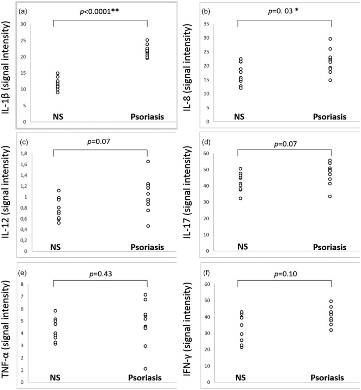
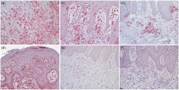
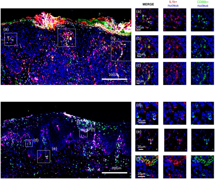
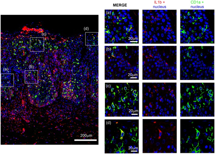
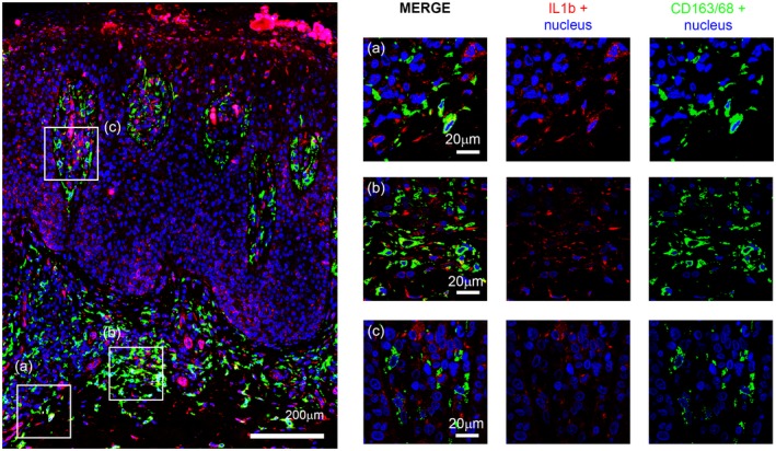
Psoriasis is a common, inflammatory immune-mediated skin disease mainly presenting with plaques whose pathogenesis is based on the central role of the interleukin (IL)-23/IL-17 axis. However, the mechanisms acting in papular lesions of early-phase psoriasis are not fully understood. The aim of this study was to assess the involvement of autoinflammation, a state of sterile inflammation mainly driven by IL-1 over-production that has been recently hypothesized to act in the early phase of disease. Lesional skin of 10 patients with recent onset, untreated psoriasis has been investigated for expression of IL-1β, IL-17, IL-23 and other cytokines involved in the disease in comparison with normal skin of 10 healthy controls using a protein array method. Immunohistochemical phenotyping of inflammatory infiltrate and co-localization experiments with immunofluorescence confocal microscopy were conducted. IL-1β was significantly more expressed in psoriasis than in normal skin (P < 0·0001). The chemokine IL-8 was also over-expressed in psoriasis (P = 0·03) while IL-12, IL-17, IL-23, tumour necrosis factor-α and interferon-γ were only slightly more expressed in psoriasis than in normal skin, without reaching statistical significance. The inflammatory infiltrate consisted mainly of neutrophils with a relevant number of macrophages and dendritic cells and only scattered, predominantly T helper 1 lymphocytes. IL-1β co-localized mainly with CD66b, a neutrophil marker, suggesting that neutrophils were the major source of this cytokine. IL-1β over-expression in combination with low expression of cytokines that are predominant in late-phase plaque psoriasis may support the role of autoinflammation in early-phase disease, possibly paving the way to randomized trials with IL-1 antagonists.
Keywords: autoinflammatory disease; cytokines; neutrophils; skin.
© 2019 British Society for Immunology.
Conflict of interest statement
None to declare.
Figures
Similar articles
-
Neutrophil exosomes enhance the skin autoinflammation in generalized pustular psoriasis via activating keratinocytes.FASEB J. 2019 Jun;33(6):6813-6828. doi: 10.1096/fj.201802090RR. Epub 2019 Feb 27. FASEB J. 2019. PMID: 30811955
-
Increased expression of IL-12p70 and IL-23 by multiple dendritic cell and macrophage subsets in plaque psoriasis.J Dermatol Sci. 2009 May;54(2):99-105. doi: 10.1016/j.jdermsci.2009.01.003. Epub 2009 Mar 4. J Dermatol Sci. 2009. PMID: 19264456
-
Keratinocyte-Derived IL-17E Contributes to Inflammation in Psoriasis.J Invest Dermatol. 2016 Oct;136(10):1970-1980. doi: 10.1016/j.jid.2016.06.009. Epub 2016 Jun 18. J Invest Dermatol. 2016. PMID: 27329229
-
Etiology and Pathogenesis of Psoriasis.Rheum Dis Clin North Am. 2015 Nov;41(4):665-75. doi: 10.1016/j.rdc.2015.07.013. Epub 2015 Sep 5. Rheum Dis Clin North Am. 2015. PMID: 26476225 Review.
-
Immunopathogenesis and role of T cells in psoriasis.Clin Dermatol. 2007 Nov-Dec;25(6):574-80. doi: 10.1016/j.clindermatol.2007.08.012. Clin Dermatol. 2007. PMID: 18021895 Review.
Cited by
-
Integrated metabolomic analysis and cytokine profiling define clusters of immuno-metabolic correlation in new-onset psoriasis.Sci Rep. 2021 May 18;11(1):10472. doi: 10.1038/s41598-021-89925-7. Sci Rep. 2021. PMID: 34006909 Free PMC article.
-
Early Neutrophil Activation in Psoriatic Skin at Relapse Following Dead Sea Climatotherapy.Exp Dermatol. 2025 Apr;34(4):e70094. doi: 10.1111/exd.70094. Exp Dermatol. 2025. PMID: 40181552 Free PMC article.
-
The Intriguing Links between Psoriasis and Bullous Pemphigoid.J Clin Med. 2022 Dec 31;12(1):328. doi: 10.3390/jcm12010328. J Clin Med. 2022. PMID: 36615129 Free PMC article. Review.
-
Skin Barrier Dysregulation in Psoriasis.Int J Mol Sci. 2021 Oct 7;22(19):10841. doi: 10.3390/ijms221910841. Int J Mol Sci. 2021. PMID: 34639182 Free PMC article. Review.
-
An update on genetic basis of generalized pustular psoriasis (Review).Int J Mol Med. 2021 Jun;47(6):118. doi: 10.3892/ijmm.2021.4951. Epub 2021 May 6. Int J Mol Med. 2021. PMID: 33955502 Free PMC article. Review.
References
-
- Nestle FO, Kaplan DH, Barker J. Psoriasis. N Engl J Med 2009; 361:496–509. - PubMed
-
- Deng Y, Chang C, Lu Q. The inflammatory response in psoriasis: a comprehensive review. Clin Rev Allergy Immunol 2016; 50:377–89. - PubMed
-
- Griffiths CE, Christophers E, Barker JN et al A classification of psoriasis vulgaris according to phenotype. Br J Dermatol 2007; 156:258–62. - PubMed
