Review
. 2020 Feb;29(2):378-390.
doi: 10.1002/pro.3725. Epub 2019 Oct 7.

Structural and functional analysis of the Hsp70/Hsp40 chaperone system

Affiliations
Review

Structural and functional analysis of the Hsp70/Hsp40 chaperone system

Qinglian Liu et al. Protein Sci. 2020 Feb.

Abstract

As one of the most abundant and highly conserved molecular chaperones, the 70-kDa heat shock proteins (Hsp70s) play a key role in maintaining cellular protein homeostasis (proteostasis), one of the most fundamental tasks for every living organism. In this role, Hsp70s are inextricably linked to many human diseases, most notably cancers and neurodegenerative diseases, and are increasingly recognized as important drug targets for developing novel therapeutics for these diseases. Hsp40s are a class of essential and universal partners for Hsp70s in almost all aspects of proteostasis. Thus, Hsp70s and Hsp40s together constitute one of the most important chaperone systems across all kingdoms of life. In recent years, we have witnessed significant progress in understanding the molecular mechanism of this chaperone system through structural and functional analysis. This review will focus on this recent progress, mainly from a structural perspective.

Keywords: Hsp40; Hsp70; molecular chaperone; neurodegenerative diseases; protein folding; proteostasis.


Conflict of interest statement

The authors declare no potential conflict of interest.

Figures

Figure 1
Figure 1
Domain organization and isolated domain structures of Hsp70s. (a) The domain organization of Hsp70s consisting of the nucleotide binding domain (NBD) and the substrate binding domain (SBD). (b) The isolated NBD structure in complex with ADP from bovine Hsc70 (PDB code: 1HPM). The bound ADP is shown as sticks. (c) The isolated SBD structure from DnaK (PDB code: 1DKX). Coloring is NBD (blue), interdomain linker (purple), SBDβ (green), SBDα (red), and the bound NR peptide (cyan). L stands for loop
Figure 2
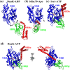
Figure 2
Representatives of full‐length structures of Hsp70s. (a) NMR positioning of DnaK NBD and SBD in a ADP‐bound state (PDB code: 2KHO). (b) The bHsc70 structure in a nucleotide‐free state (PDB code: 1YUW). (c) The structure of Sse1 in complex with ATP (PDB code: 2QXL). (d) The structure of DnaK in complex with ATP solved by our group (PDB code: 4JNE). Coloring is NBD (blue), interdomain linker (purple), SBDβ (green), and SBDα (red). ATP molecules are shown as sticks
Figure 3
Figure 3
Conformations of the polypeptide‐binding pocket. The SBDβ structures from the isolated SBD of DnaK (a, PDB code: 1DKX), DnaK‐ATP solved by our group (b, PDB code: 4JNE), DnaK‐ATP solved by Mayer's group (c, PDB code: 4B9Q), BiP‐ATP2 (d, PDB code: 8ASY). For a, b, and c, the structures are aligned based on the Cα atoms of the L1,2 and L3,4. The BiP‐ATP2 structure (d) is aligned with the DnaK‐ATP structure (b) based on the Cα atoms of the β3‐β4 region
Figure 4
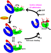
Figure 4
Schematic of the proposed Hsp70 chaperone cycle. Hsp70 domain color assignments are as in Figure 1a. Polypeptide substrate is highlighted in black
Figure 5
Figure 5
Hsp40 domain organization and structures. (a) Domain organization of Hsp40s (JDP). J, J‐domain; G/F, G/F rich domain; CTD, C‐terminal domain; ZFLR, zinc finger‐like region; D, dimerization domain. (b) Ribbon diagram of the J‐domain structure from the E. coli Hsp40 DnaJ (PDB code: 1XBL). (c) The CTD structure of a yeast Hsp40 Ydj1 (PDB code: 1NLT). The domains are labeled. The bound peptide substrate is highlighted in red, and the two associated Zn ions are colored in cyan. (d) The structural model of full‐length DnaJ from Thermus thermophiles (PDB code: 4 J80). The J‐domain is highlighted in orange, and the HPD motif is shown as blue sticks
Figure 6
Figure 6
Structures of the Hsp70‐Hsp40 complexes. (a) The structure of bHsc70 NBD (blue) crosslinked with the J‐domain of Auxillin (orange) (PDB code: 2QWO). (b) The structure of DnaK fused to the DnaJ's J‐domain (PDB code: 5NRO). The domain coloring of DnaK is NBD (blue), interdomain linker (purple), SBDβ (green), and SBDα (red). The J‐domain of DnaJ is in orange

Similar articles

Cited by

References

    1. Mayer MP, Bukau B. Hsp70 chaperones: Cellular functions and molecular mechanism. Cell Mol Life Sci. 2005;62:670–684. - PMC - PubMed
    1. Hartl FU, Hayer‐Hartl M. Converging concepts of protein folding in vitro and in vivo. Nat Struct Mol Biol. 2009;16:574–581. - PubMed
    1. Bukau B, Weissman J, Horwich A. Molecular chaperones and protein quality control. Cell. 2006;125:443–451. - PubMed
    1. Bukau B, Deuerling E, Pfund C, Craig EA. Getting newly synthesized proteins into shape. Cell. 2000;101:119–122. - PubMed
    1. Bukau B, Horwich AL. The Hsp70 and Hsp60 chaperone machines. Cell. 1998;92:351–366. - PubMed

Publication types

MeSH terms

Substances