. 2019 Nov 1;317(5):L717-L736.
doi: 10.1152/ajplung.00132.2019. Epub 2019 Sep 11.

Clinically relevant model of pneumococcal pneumonia, ARDS, and nonpulmonary organ dysfunction in mice

Affiliations

Clinically relevant model of pneumococcal pneumonia, ARDS, and nonpulmonary organ dysfunction in mice

Jeffrey E Gotts et al. Am J Physiol Lung Cell Mol Physiol. .

Abstract

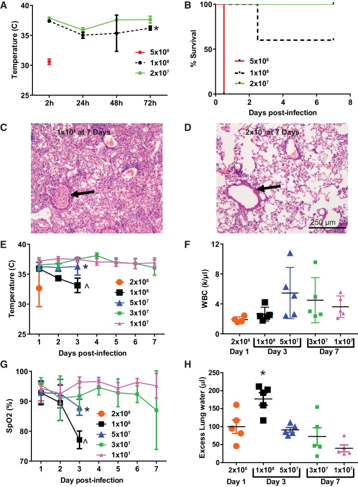
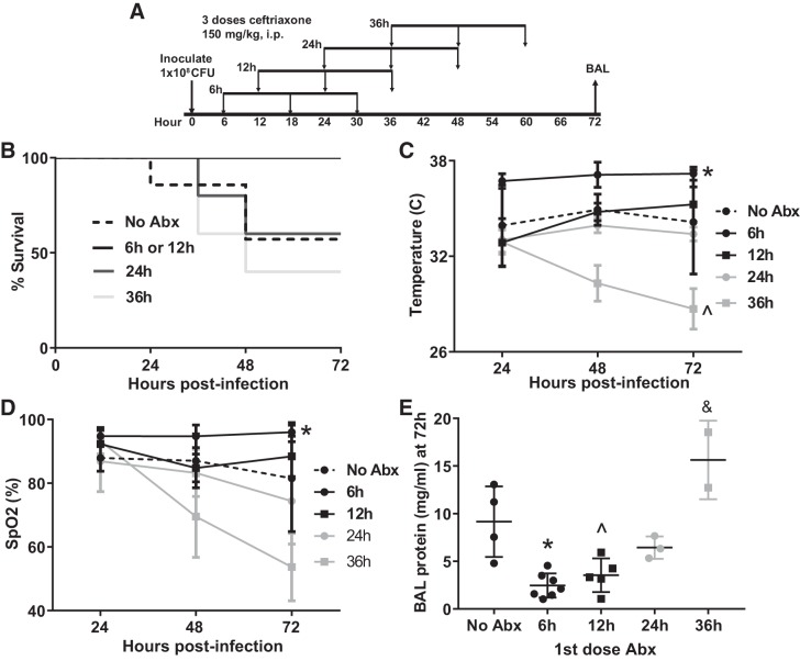
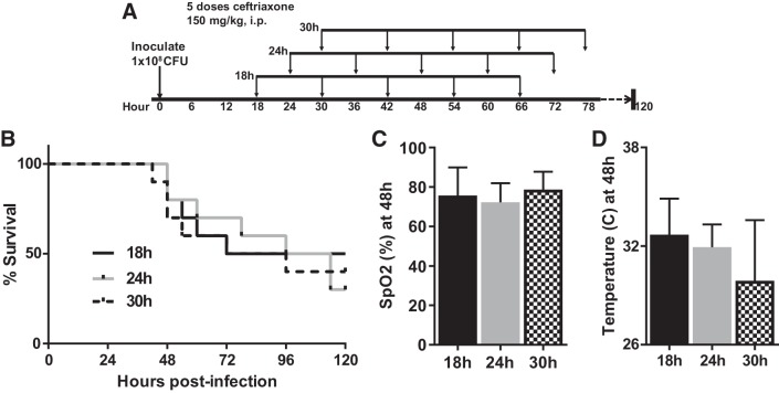
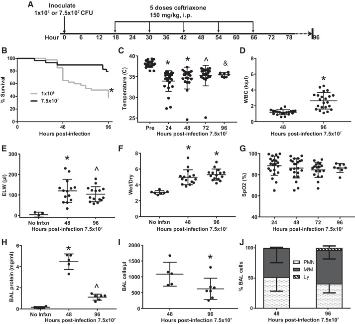
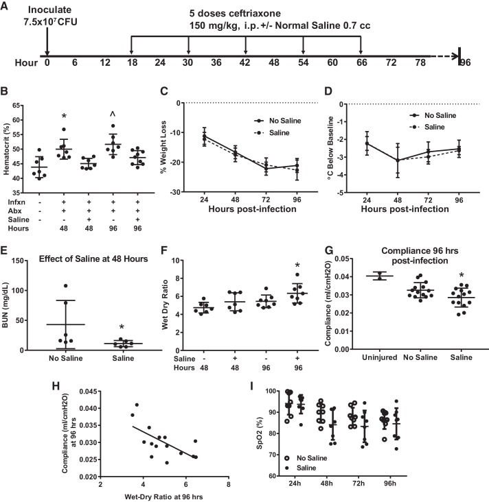
Pneumonia is responsible for more deaths in the United States than any other infectious disease. Severe pneumonia is a common cause of acute respiratory failure and acute respiratory distress syndrome (ARDS). Despite the introduction of effective antibiotics and intensive supportive care in the 20th century, death rates from community-acquired pneumonia among patients in the intensive care unit remain as high as 35%. Beyond antimicrobial treatment, no targeted molecular therapies have yet proven effective, highlighting the need for additional research. Despite some limitations, small animal models of pneumonia and the mechanistic insights they produce are likely to continue to play an important role in generating new therapeutic targets. Here we describe the development of an innovative mouse model of pneumococcal pneumonia developed for enhanced clinical relevance. We first reviewed the literature of small animal models of bacterial pneumonia that incorporated antibiotics. We then did a series of experiments in mice in which we systematically varied the pneumococcal inoculum and the timing of antibiotics while measuring systemic and lung-specific end points, producing a range of models that mirrors the spectrum of pneumococcal lung disease in patients, from mild self-resolving infection to severe pneumonia refractory to antibiotics. A delay in antibiotic treatment resulted in ongoing inflammation and renal and hepatic dysfunction despite effective bacterial killing. The addition of fluid resuscitation to the model improved renal function but worsened the severity of lung injury based on direct measurements of pulmonary edema and lung compliance, analogous to patients with pneumonia and sepsis who develop ARDS following fluid administration.

Keywords: ARDS; acute lung injury; pneumococcus; pneumonia; sepsis.


Conflict of interest statement

No conflicts of interest, financial or otherwise, are declared by the authors.

Figures

Fig. 1.
Fig. 1.
Streptococcus pneumoniae dose response. A: mice inoculated intranasally with between 2 × 10⁷ and 5 × 10⁸ colony-forming units (CFU) of S. pneumoniae developed hypothermia. Data are mean ± SD; n = 5 per dosing group. *P = 0.0025 compared with 2 × 10⁷ by repeated measures ANOVA. B: S. pneumoniae caused between 0% and 100% mortality across this inoculation range; P = 0.0004 for log rank test for trend. C: histology (hematoxylin-eosin) at 7 days in survivors of 1 × 10⁸ CFU reveals severe lobar pneumonia with airway occlusion by inflammatory exudate (arrow). D: at lower doses, airways were consistently patent (arrow), and a more modest inflammatory infiltrate was observed. E: S. pneumoniae causes hypothermia at doses greater than 3 × 10⁷ CFU. *P = 0.0007, ^P < 0.0001 compared with 1 × 10⁷ over the first 3 days postinfection by Dunnett’s multiple comparisons test. F: leukopenia was most pronounced at early time points and doses greater than 5 × 10⁷ CFU. P = 0.16 for ANOVA. G: arterial oxygen saturation (SpO2) tended to decline in a dose-dependent manner for the first 72 h postinfection. *P = 0.04, ^P < 0.006 compared with 1 × 10⁷ over the first 3 days postinfection by Dunnett’s multiple comparisons test. H: pulmonary edema as measured by excess extravascular lung water (in the interstitial and alveolar spaces) mirrored the changes in oxygenation. Overall ANOVA P = 0.0002; *P = 0.03 compared with 2 × 10⁸, P = 0.01 compared with 5 × 10⁷, P = 0.002 compared with 3 × 10⁷, P < 0.0001 compared with 1 × 10⁷ by Tukey’s multiple comparisons test. WBC, white blood cell.
Fig. 2.
Fig. 2.
Influence of timing of first dose of antibiotics on the model. A: schematic depicting experimental procedures. Mice were inoculated with 1 × 10⁸ colony-forming units (CFU) Streptococcus pneumoniae followed by 3 doses of ceftriaxone beginning at 6, 12, 24, or 36 h postinfection. B: there was a trend toward decreased survival as antibiotics (Abx) were delayed (n = 5–7 mice/group). P = 0.07 by log rank test. C: mice treated with antibiotics beginning at 6 h were significantly less hypothermic than untreated mice. Surprisingly, initiation of antibiotics 36 h postinfection caused more severe hypothermia than was observed without antibiotics. *P = 0.0005, ^P = 0.005 compared with no antibiotics by Dunnett’s multiple comparisons test. D: arterial hypoxemia was most severe in mice treated beginning 36 h postinfection. *P = 0.009 compared with no Abx by Dunnett’s multiple comparisons test. Significant mortality in the 36-h group reduced statistical power at 72 h. SpO2, arterial oxygen saturation. E: bronchoalveolar lavage (BAL) protein mirrored oxygenation with a 36-h delay in antibiotics causing higher BAL protein 72 h postinfection than in untreated mice. *P = 0.0009, ^P = 0.007, &P = 0.016 compared with no Abx by Dunnett’s multiple comparisons test.
Fig. 3.
Fig. 3.
Fine-tuning the antibiotic regimen to optimize 7-day survival. A: schematic depicting experimental procedures. Mice were inoculated with 1 × 10⁸ colony-forming units (CFU) Streptococcus pneumoniae and then received 5 doses of ceftriaxone beginning 18, 24, or 30 h later. B: mice administered the first dose of antibiotics 18 h postinfection had 50% survival at 7 days. n = 10 mice per group. P = 0.23 for log rank test for trend. C: arterial oxygen saturation (SpO2) was similar across the 3 cohorts. Data are mean ± SD; P = 0.53 by ANOVA. D: body temperature trended lower with progressive delays in initiation of antibiotics. P = 0.11 by Kruskal–Wallis test.
Fig. 4.
Fig. 4.
Reducing the bacterial inoculum to improve 96-h survival while maintaining a moderately severe lung injury. A: schematic depicting experimental procedures. Mice were inoculated with either 1 × 10⁸ or 7.5 × 10⁷ colony-forming units (CFU) Streptococcus pneumoniae and then treated with 5 doses of ceftriaxone beginning 18 h postinfection. B: survival was ~80% at 96 h in the lower-dose group (*by log rank, n = 29–40 per group). C: mice were hypothermic between 24 and 96 h postinfection. *P < 0.0001, ^P = 0.0001, &P = 0.01 by Dunnett’s multiple comparisons test. D: peripheral leukopenia resolves between 48 and 96 h postinfection. *P < 0.0001 by unpaired t test. E: excess lung water (ELW), a measure of edema in the interstitial and alveolar spaces, remained elevated above baseline at 48 and 96 h postinfection. *P = 0.0024, ^P = 0.0099 by Dunn’s multiple comparisons test. F: wet-dry ratio was similarly increased at 48 and 96 h postinfection. *P < 0.0001 by Dunnett’s multiple comparisons test. G: mean arterial oxygen saturation (SpO2) remained in the high 80s during the entire 96 h postinfection, though the standard deviation was high. P = 0.33 by Kruskal–Wallis. H: bronchoalveolar lavage (BAL) protein peaked at 48 h postinfection and remained significantly above baseline at 96 h. *P < 0.0001 compared with no infection (No Infxn) and 96 h, ^P = 0.0194 compared with No Infxn by Tukey’s multiple comparisons test. I: BAL cellularity declined significantly between 48 and 96 h postinfection; *P = 0.0303 by Mann–Whitney. J: composition of BAL cellularity was mostly neutrophils and monocyte/macrophages at both time points, though there was a trend for an increase in lymphocytes between 48 and 96 h postinfection, from 0.5% to 3.7%, P = 0.12 by Mann–Whitney. WBC, white blood cell.
Fig. 5.
Fig. 5.
Histological evidence of lung injury. AH: representative photomicrographs from the lungs of mice inoculated with 7.5 × 10⁷ colony-forming units (CFU) Streptococcus pneumoniae followed by ceftriaxone beginning 18 h later. Alveolar septal thickness (asterisks) is greatest at 48 h without antibiotics (Abx), but it remains abnormal at 48 and 96 h in antibiotic-treated mice. Inflammation is primarily neutrophilic at 48 h with or without antibiotics (arrows); by 96 h postinfection, increasing numbers of monocytic and lymphocytic cells are identified (arrowheads). No Infxn, no infection.
Fig. 6.
Fig. 6.
Bronchoalveolar lavage (BAL) cytokine expression. A: level of KC (murine homologue of IL-8, a potent neutrophil chemokine) in BAL at 48 h was significantly reduced with antibiotic (Abx) treatment but was still elevated above noninfected controls. By 96 h, BAL KC had returned to baseline in the antibiotic-treated model. *P = 0.0044 compared with −Infxn, −Abx; ^P = 0.03 compared with +Infxn −Abx by Dunn’s multiple comparisons test. B: BAL MIP-1α/CCL3 was also higher in untreated infected mice at 48 h and declined substantially by 96 h but remained elevated relative to uninfected mice. P < 0.004 for all comparisons by Tukey’s multiple comparisons test. C: monocyte chemokine MCP-1/CCL2 was markedly elevated in BAL 48 h postinfection but did not differ with respect to antibiotics (P < 0.0001 for both injured groups compared with uninfected mice by Tukey’s multiple comparisons test). By 96 h, levels declined by 2 orders of magnitude but remained slightly elevated above baseline (P < 0.0001). D: similarly, the monocyte chemokine IP-10/CXCL10 was elevated at 48 h regardless of antibiotics (P < 0.0001 for both injured groups compared with uninfected mice, by Tukey’s multiple comparisons test), declining rapidly by 96 h. E: BAL TNF-α was significantly higher without antibiotic treatment at 48 h but rapidly declined to baseline by 96 h. *P < 0.0001 compared with all other groups by Tukey’s multiple comparisons test. F: in contrast, BAL IL-6 was not significantly different at 48 h with respect to antibiotic therapy (P < 0.0001 for both infected groups at 48 h relative to uninfected groups by Tukey’s multiple comparisons test). Abx, antibiotics; Infxn, infection.
Fig. 7.
Fig. 7.
Persistent hypothermia and systemic inflammation despite bacterial clearance. A: schematic depicting experimental procedures. Mice were inoculated with 7.5 × 10⁷ colony-forming units (CFU) Streptococcus pneumoniae and then treated with 5 doses of ceftriaxone beginning 18 h postinfection. Mice were euthanized at 96 or 168 h (4 or 7 days) postinfection. B: persistent hypothermia depicted as mean departure from preinfection temperature (± SD), n = 8–24 mice per time point. P = 0.24 by ANOVA. C: bacteremia resolved within 48 h in antibiotic-treated mice. Blood cultures remained negative 7 days postinfection, more than 4 days after the completion of antibiotics. *P = 0.0015 compared with 48 h, P = 0.0004 compared with 96 h, P = 0.0002 compared with 168 h, by Dunn’s multiple comparisons test. D: airspace pneumococci were cleared by 48 h postinfection and remained sterile at 96 h. About half of the mice had low levels of detectable bacteria 7 days postinfection, though these did not appear to be pneumococci based on the absence of hemolysis. *P = 0.003 compared with 48 h, P < 0.0001 compared with 96 h, P = 0.003 compared with 168 h, by Dunn’s multiple comparisons test. E: plasma IL-6 was significantly elevated 48 h postinfection (*P = 0.04 compared with no infection, P = 0.04 compared with 96 h, by Tukey’s multiple comparisons test). F: plasma TNF-α was significantly increased 48 h postinfection (*P = 0.025 compared with no infection, by Tukey’s multiple comparisons test). G: plasma IL-1β was elevated at 48 h (*P = 0.001 compared with no infection, P = 0.0003 compared with 96 h, by Tukey’s multiple comparisons test). Abx, antibiotics; BAL, bronchoalveolar lavage; Infxn, infection.
Fig. 8.
Fig. 8.
Evidence of renal and hepatic dysfunction. A: Schematic depicting experimental procedures. Mice were inoculated with 7.5 × 10⁷ colony-forming units (CFU) Streptococcus pneumoniae and then treated with 5 doses of ceftriaxone beginning 18 h postinfection followed by euthanization at 48 or 96 h. B: level of blood urea nitrogen (BUN) was increased above baseline at 48 h postinfection, declining back to baseline by 96 h; *P = 0.05 compared with no infection (No Infxn), P = 0.009 compared with 96 h by Dunn’s multiple comparisons test. C: alanine aminotransferase (ALT) was significantly increased above baseline at 48 and 96 h postinfection. *P = 0.0004 compared with 48 h, P = 0.001 compared with 96 h by Tukey’s multiple comparisons test. D and F: hematoxylin-eosin (H&E)-stained sections of renal cortex reveals grossly normal glomeruli and tubules 48 h postinfection. E and G: H&E stained sections of liver parenchyma 48 h postinfection show no evidence of significant centrilobular necrosis or other histologic abnormalities.
Fig. 9.
Fig. 9.
Fluid resuscitation increases lung edema and worsens lung compliance. A: schematic depicting experimental procedures. Mice were inoculated with 7.5 × 10⁷ colony-forming units (CFU) Streptococcus pneumoniae and then treated with 5 doses of ceftriaxone beginning 18 h postinfection and then every 12 h × 5, with euthanization at 48 or 96 h. Half of the mice also received 700 μL ip of normal saline with each dose of antibiotics. B: saline treatment reversed hemoconcentration at 48 and 96 h, *P = 0.0046 vs. uninfected, P = 0.03 vs. saline at 48 h, by Tukey’s multiple comparisons test; ^P = 0.0003 vs. uninfected, P = 0.037 vs. saline at 96 h. C and D: saline administration did not significantly affect weight loss or hypothermia. Not significant by repeated measures ANOVA. E: mice treated with saline had normalized blood urea nitrogen (BUN), consistent with correction of renal hypoperfusion; *P = 0.026 by Mann–Whitney. F: saline treatment compared with no saline increased lung edema at 96 h as measured by wet-dry ratio. *P = 0 0.05 by Mann–Whitney. G: lung dynamic compliance at 96 h was significantly reduced by saline administration, *P = 0.047 by unpaired t test. H: lung compliance and wet-dry ratio were inversely correlated, Spearman r = −0.62, P = 0.02. I: there was a trend for lower arterial oxygenation between 24 and 96 h in fluid-resuscitated mice. SpO2, arterial oxygen saturation. P = 0.12 for group effect in repeated measures ANOVA. Abx, antibiotics; Infxn, infection.

Similar articles

Cited by

References

    1. Abdulnour RE, Sham HP, Douda DN, Colas RA, Dalli J, Bai Y, Ai X, Serhan CN, Levy BD. Aspirin-triggered resolvin D1 is produced during self-resolving gram-negative bacterial pneumonia and regulates host immune responses for the resolution of lung inflammation. Mucosal Immunol 9: 1278–1287, 2016. doi:10.1038/mi.2015.129. - DOI - PMC - PubMed
    1. Ansfield MJ, Woods DE, Johanson WG Jr. Lung bacterial clearance in murine pneumococcal pneumonia. Infect Immun 17: 195–204, 1977. - PMC - PubMed
    1. Bansal S, Chhibber S. Curcumin alone and in combination with augmentin protects against pulmonary inflammation and acute lung injury generated during Klebsiella pneumoniae B5055-induced lung infection in BALB/c mice. J Med Microbiol 59: 429–437, 2010. doi:10.1099/jmm.0.016873-0. - DOI - PubMed
    1. Bansal S, Chhibber S. Phytochemical-induced reduction of pulmonary inflammation during Klebsiella pneumoniae lung infection in mice. J Infect Dev Ctries 8: 838–844, 2014. doi:10.3855/jidc.3277. - DOI - PubMed
    1. Bast DJ, Yue M, Chen X, Bell D, Dresser L, Saskin R, Mandell LA, Low DE, de Azavedo JC. Novel murine model of pneumococcal pneumonia: use of temperature as a measure of disease severity to compare the efficacies of moxifloxacin and levofloxacin. Antimicrob Agents Chemother 48: 3343–3348, 2004. doi:10.1128/AAC.48.9.3343-3348.2004. - DOI - PMC - PubMed

Publication types

MeSH terms

Substances

LinkOut - more resources