

Hemoglobin alters vitamin carrier uptake and vitamin D metabolism in proximal tubule cells: implications for sickle cell disease
- PMID: 31509446
- PMCID: PMC6879883
- DOI: 10.1152/ajpcell.00287.2019
Hemoglobin alters vitamin carrier uptake and vitamin D metabolism in proximal tubule cells: implications for sickle cell disease
Abstract
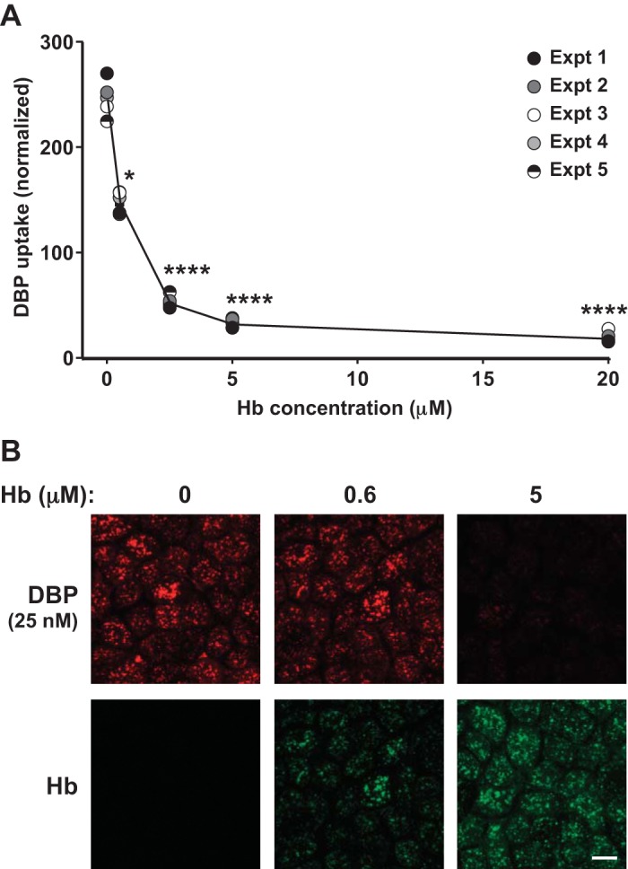
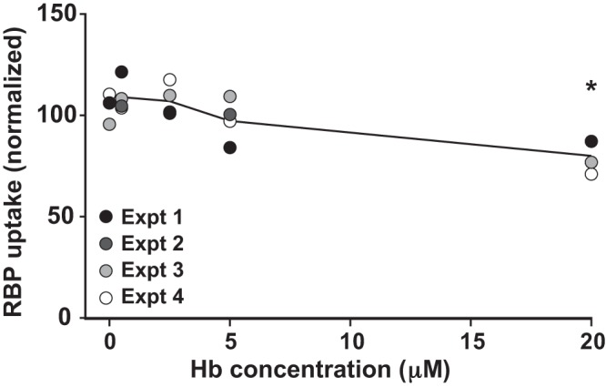
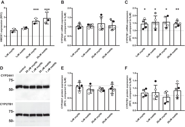
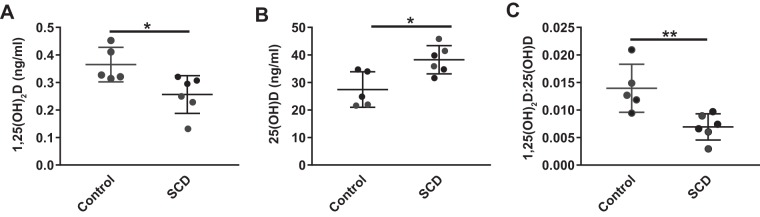
Kidney disease, including proximal tubule (PT) dysfunction, and vitamin D deficiency are among the most prevalent complications in sickle cell disease (SCD) patients. Although these two comorbidities have never been linked in SCD, the PT is the primary site for activation of vitamin D. Precursor 25-hydroxyvitamin D [25(OH)D] bound to vitamin D-binding protein (DBP) is taken up by PT cells via megalin/cubilin receptors, hydroxylated to the active 1,25-dihydroxyvitamin D [1,25(OH)2D] form, and released into the bloodstream. We tested the hypothesis that cell-free hemoglobin (Hb) filtered into the PT lumen impairs vitamin D uptake and metabolism. Hb at concentrations expected to be chronically present in the ultrafiltrate of SCD patients competed directly with DBP for apical uptake by PT cells. By contrast, uptake of retinol binding protein was impaired only at considerably higher Hb concentrations. Prolonged exposure to Hb led to increased oxidative stress in PT cells and to a selective increase in mRNA levels of the CYP27B1 hydroxylase, although protein levels were unchanged. Hb exposure also impaired vitamin D metabolism in PT cells, resulting in reduced ratio of 1,25(OH)2D:25(OH)D. Moreover, plasma levels of 1,25(OH)2D were reduced in a mouse model of SCD. Together, our data suggest that Hb released by chronic hemolysis has multiple effects on PT function that contribute to vitamin D deficiency in SCD patients.
Keywords: endocytosis; megalin; proximal tubule; sickle cell disease; vitamin D.
Conflict of interest statement
No conflicts of interest, financial or otherwise, are declared by the authors.
Figures

Similar articles
-
Hemoglobin inhibits albumin uptake by proximal tubule cells: implications for sickle cell disease.Am J Physiol Cell Physiol. 2017 Jun 1;312(6):C733-C740. doi: 10.1152/ajpcell.00021.2017. Epub 2017 Mar 29. Am J Physiol Cell Physiol. 2017. PMID: 28356267 Free PMC article.
-
Proximal tubule endocytic apparatus as the specific renal uptake mechanism for vitamin D-binding protein/25-(OH)D3 complex.Nephrology (Carlton). 2006 Dec;11(6):510-5. doi: 10.1111/j.1440-1797.2006.00704.x. Nephrology (Carlton). 2006. PMID: 17199789 Review.
-
Cubilin- and megalin-mediated uptake of albumin in cultured proximal tubule cells of opossum kidney.Kidney Int. 2000 Oct;58(4):1523-33. doi: 10.1046/j.1523-1755.2000.00314.x. Kidney Int. 2000. PMID: 11012887
-
Interactions of vitamin D and the proximal tubule.Pediatr Nephrol. 2016 Jan;31(1):7-14. doi: 10.1007/s00467-015-3050-5. Epub 2015 Jan 25. Pediatr Nephrol. 2016. PMID: 25618772 Review.
-
The vitamin D receptor in the proximal renal tubule is a key regulator of serum 1α,25-dihydroxyvitamin D₃.Am J Physiol Endocrinol Metab. 2015 Feb 1;308(3):E201-5. doi: 10.1152/ajpendo.00422.2014. Epub 2014 Nov 25. Am J Physiol Endocrinol Metab. 2015. PMID: 25425001
Cited by
-
Hypothesis: Low Vitamin A and D Levels Worsen Clinical Outcomes When Children with Sickle Cell Disease Encounter Parvovirus B19.Nutrients. 2022 Aug 19;14(16):3415. doi: 10.3390/nu14163415. Nutrients. 2022. PMID: 36014920 Free PMC article.
-
The Worst Things in Life are Free: The Role of Free Heme in Sickle Cell Disease.Front Immunol. 2021 Jan 27;11:561917. doi: 10.3389/fimmu.2020.561917. eCollection 2020. Front Immunol. 2021. PMID: 33584641 Free PMC article. Review.
-
Influence of hydroxyurea on tubular phosphate handling in sickle cell nephropathy.Hematol Transfus Cell Ther. 2024 Nov;46 Suppl 5(Suppl 5):S163-S169. doi: 10.1016/j.htct.2023.11.015. Epub 2024 Feb 14. Hematol Transfus Cell Ther. 2024. PMID: 38485550 Free PMC article.
-
Functional characterization of nutraceuticals using spectral clustering: Centrality of caveolae-mediated endocytosis for management of nitric oxide and vitamin D deficiencies and atherosclerosis.Front Nutr. 2022 Aug 15;9:885364. doi: 10.3389/fnut.2022.885364. eCollection 2022. Front Nutr. 2022. PMID: 36046126 Free PMC article.
-
Vitamin D level, lipid profile, and vitamin D receptor and transporter gene variants in sickle cell disease patients from Kurdistan of Iraq.J Clin Lab Anal. 2021 Sep;35(9):e23908. doi: 10.1002/jcla.23908. Epub 2021 Jul 14. J Clin Lab Anal. 2021. PMID: 34261187 Free PMC article.
References
Publication types
MeSH terms
Substances
Grants and funding
LinkOut - more resources
Full Text Sources
Medical
Molecular Biology Databases
Miscellaneous