

Sensory enhancement amplifies interlimb cutaneous reflexes in wrist extensor muscles
- PMID: 31509473
- PMCID: PMC6879954
- DOI: 10.1152/jn.00324.2019
Sensory enhancement amplifies interlimb cutaneous reflexes in wrist extensor muscles
Abstract
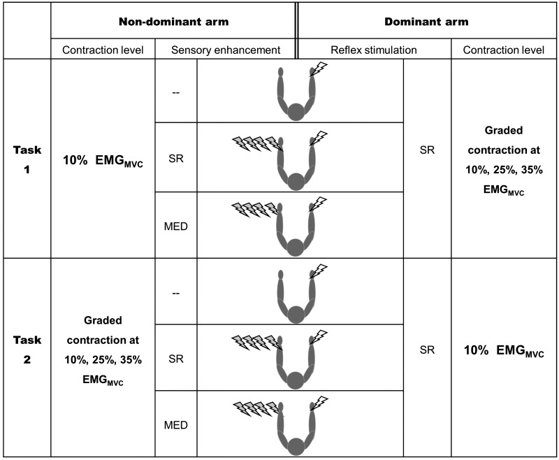
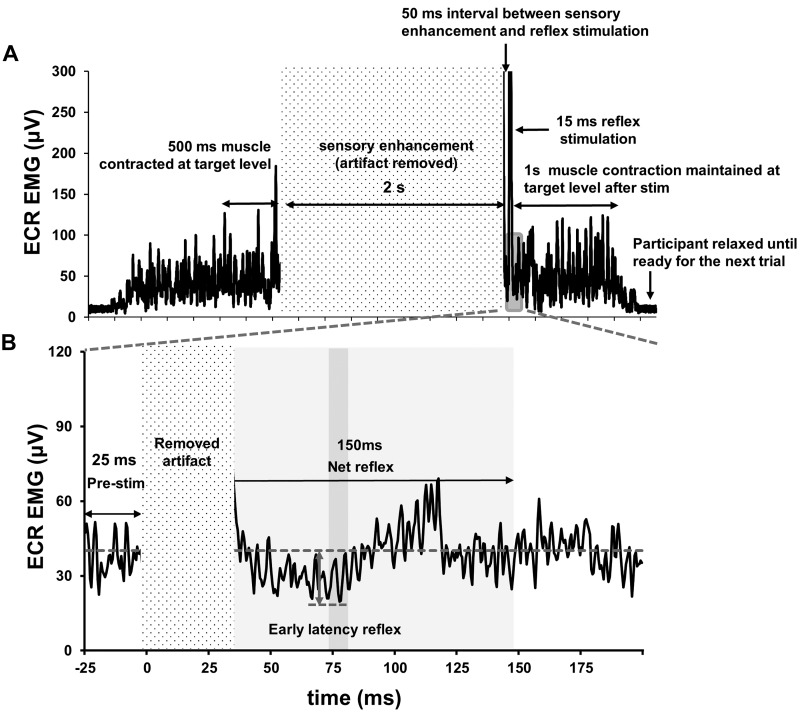
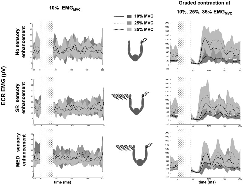
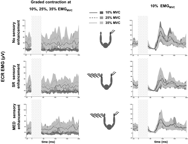
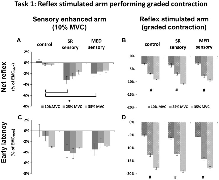
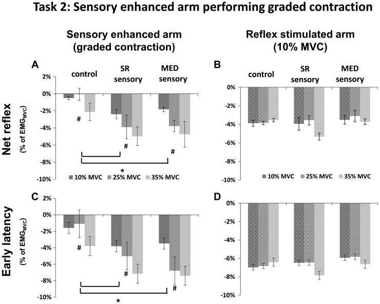
Interlimb neural connections support motor tasks such as locomotion and cross-education strength training. Somatosensory pathways that can be assessed with cutaneous reflex paradigms assist in subserving these connections. Many studies show that stimulation of cutaneous nerves elicits reflexes in muscles widespread across the body and induces neural plasticity after training. Sensory enhancement, such as long-duration trains of transcutaneous stimulation, facilitates performance during rehabilitation training or fatiguing motor tasks. Performance improvements due to sensory stimulation may be caused by altered spinal and corticospinal excitability. However, how enhanced sensory input regulates the excitability of interlimb cutaneous reflex pathways has not been studied. Our purpose was to investigate the effects of sensory enhancement on interlimb cutaneous reflexes in wrist extensor muscles. Stimulation to provide sensory enhancement (2-s trains at 150 Hz to median or superficial radial nerves) or evoke cutaneous reflexes (15-ms trains at 300 Hz to superficial radial nerve) was applied in different arms while participants (n = 13) performed graded isometric wrist extension. Wrist extensor electromyography and cutaneous reflexes were measured bilaterally. We found amplified inhibitory reflexes in the arm receiving superficial radial and median nerve sensory enhancement with net reflex amplitudes decreased by 709.5% and 695.3% repetitively. This suggests sensory input alters neuronal excitabilities in the interlimb cutaneous pathways. These findings have potential application in facilitating motor function recovery through alterations in spinal cord excitability enhancing sensory input during targeted rehabilitation and sports training.NEW & NOTEWORTHY We show that sensory enhancement increases excitability in interlimb cutaneous pathways and that these effects are not influenced by descending motor drive on the contralateral side. These findings confirm the role of sensory input and cutaneous pathways in regulating interlimb movements. In targeted motor function training or rehabilitation, sensory enhancement may be applied to facilitate outcomes.
Keywords: cutaneous reflexes; interlimb reflexes; sensory enhancement.
Conflict of interest statement
No conflicts of interest, financial or otherwise, are declared by the authors.
Figures
Similar articles
-
Effects of enhanced cutaneous sensory input on interlimb strength transfer of the wrist extensors.Physiol Rep. 2020 Mar;8(6):e14406. doi: 10.14814/phy2.14406. Physiol Rep. 2020. PMID: 32222042 Free PMC article.
-
Enhanced somatosensory feedback modulates cutaneous reflexes in arm muscles during self-triggered or prolonged stimulation.Exp Brain Res. 2020 Feb;238(2):295-304. doi: 10.1007/s00221-019-05678-w. Epub 2020 Jan 2. Exp Brain Res. 2020. PMID: 31897517
-
Effects of wrist position on reciprocal inhibition and cutaneous reflex amplitudes in forearm muscles.Neurosci Lett. 2018 Jun 11;677:37-43. doi: 10.1016/j.neulet.2018.04.031. Epub 2018 Apr 21. Neurosci Lett. 2018. PMID: 29684529
-
Contributions to the understanding of gait control.Dan Med J. 2014 Apr;61(4):B4823. Dan Med J. 2014. PMID: 24814597 Review.
-
Pushing the Limits of Interlimb Connectivity: Neuromodulation and Beyond.Biomedicines. 2025 May 19;13(5):1228. doi: 10.3390/biomedicines13051228. Biomedicines. 2025. PMID: 40427058 Free PMC article. Review.
Cited by
-
I tap myself, and you tap me: bimanual predictive and reactive grip force control as a function of age.Exp Brain Res. 2024 Nov;242(11):2613-2622. doi: 10.1007/s00221-024-06925-5. Epub 2024 Sep 25. Exp Brain Res. 2024. PMID: 39320436
-
Post-Stroke Rehabilitation: Neurophysiology Processes of Bilateral Movement Training and Interlimb Coupling-A Systematic Review.J Clin Med. 2025 May 27;14(11):3757. doi: 10.3390/jcm14113757. J Clin Med. 2025. PMID: 40507519 Free PMC article. Review.
-
Effects of enhanced cutaneous sensory input on interlimb strength transfer of the wrist extensors.Physiol Rep. 2020 Mar;8(6):e14406. doi: 10.14814/phy2.14406. Physiol Rep. 2020. PMID: 32222042 Free PMC article.
-
Nervous system modulation through electrical stimulation in companion animals.Acta Vet Scand. 2021 May 30;63(1):22. doi: 10.1186/s13028-021-00585-z. Acta Vet Scand. 2021. PMID: 34053462 Free PMC article. Review.
-
Plantarflexion force is amplified with sensory stimulation during ramping submaximal isometric contractions.J Neurophysiol. 2020 Apr 1;123(4):1427-1438. doi: 10.1152/jn.00650.2019. Epub 2020 Mar 11. J Neurophysiol. 2020. PMID: 32159422 Free PMC article.
References
Publication types
MeSH terms
LinkOut - more resources
Full Text Sources