

The intrinsically disordered region of the cytokinetic F-BAR protein Cdc15 performs a unique essential function in maintenance of cytokinetic ring integrity
- PMID: 31509478
- PMCID: PMC6789166
- DOI: 10.1091/mbc.E19-06-0314
The intrinsically disordered region of the cytokinetic F-BAR protein Cdc15 performs a unique essential function in maintenance of cytokinetic ring integrity
Abstract
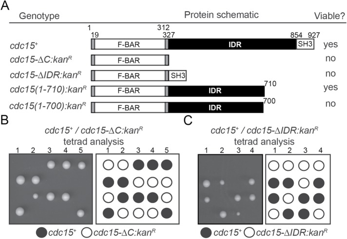
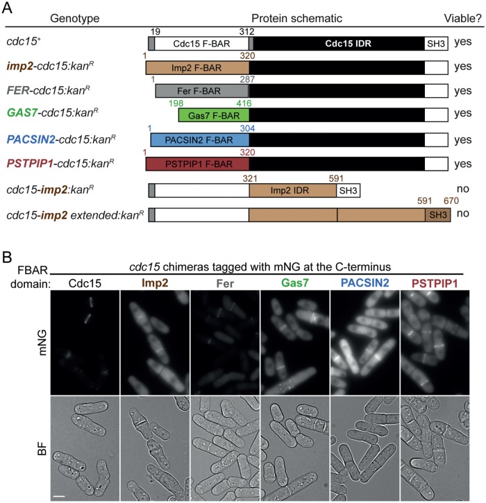
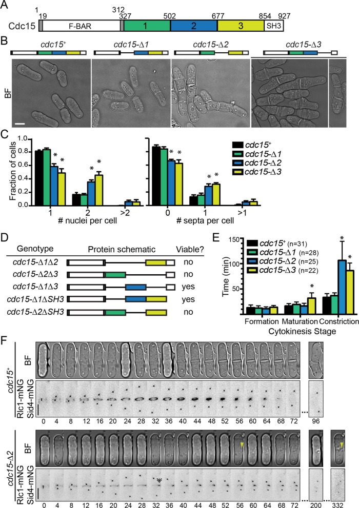
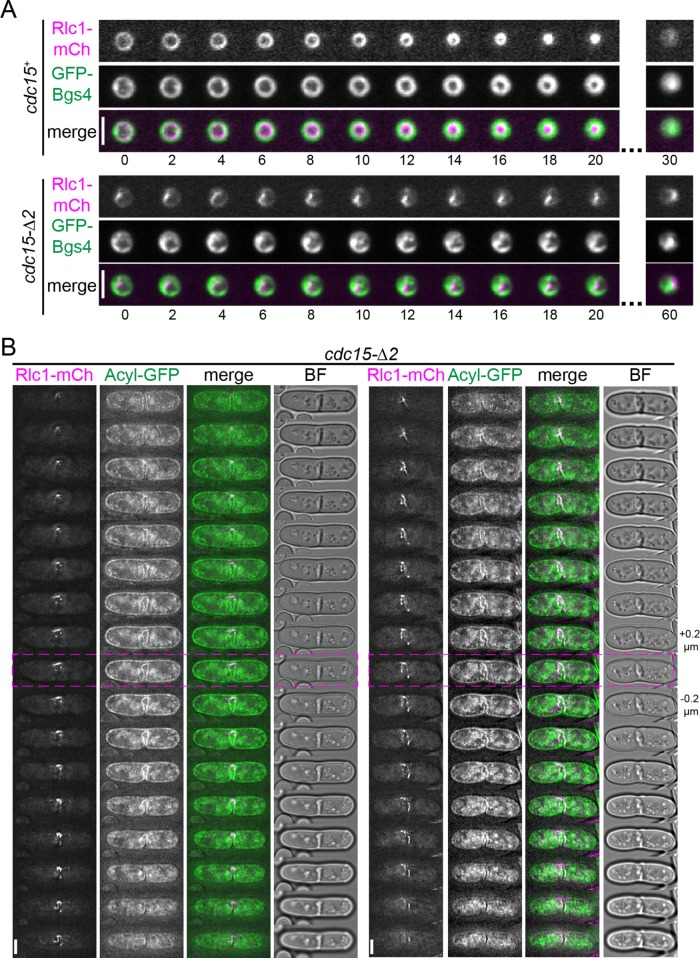
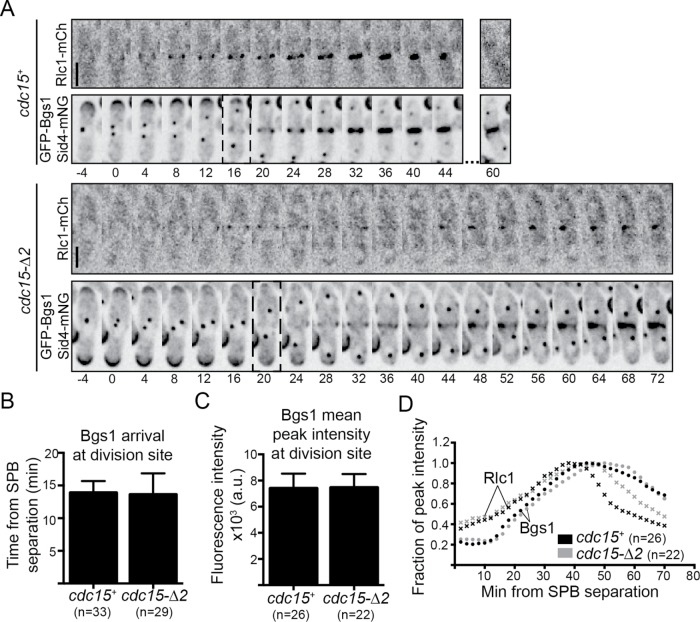
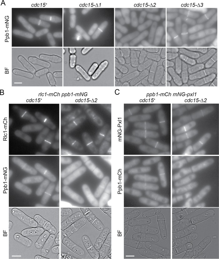
Successful separation of two daughter cells (i.e., cytokinesis) is essential for life. Many eukaryotic cells divide using a contractile apparatus called the cytokinetic ring (CR) that associates dynamically with the plasma membrane (PM) and generates force that contributes to PM ingression between daughter cells. In Schizosaccharomyces pombe, important membrane-CR scaffolds include the paralogous F-BAR proteins Cdc15 and Imp2. Their conserved protein structure consists of the archetypal F-BAR domain linked to an SH3 domain by an intrinsically disordered region (IDR). Functions have been assigned to the F-BAR and SH3 domains. In this study we probed the function of the central IDR. We found that the IDR of Cdc15 is essential for viability and cannot be replaced by that of Imp2, whereas the F-BAR domain of Cdc15 can be swapped with several different F-BAR domains, including that of Imp2. Deleting part of the IDR results in CR defects and abolishes calcineurin phosphatase localization to the CR. Together these results indicate that Cdc15's IDR has a nonredundant essential function that coordinates regulation of CR architecture.
Figures
Similar articles
-
Multiple polarity kinases inhibit phase separation of F-BAR protein Cdc15 and antagonize cytokinetic ring assembly in fission yeast.Elife. 2023 Feb 7;12:e83062. doi: 10.7554/eLife.83062. Elife. 2023. PMID: 36749320 Free PMC article.
-
Opposite Surfaces of the Cdc15 F-BAR Domain Create a Membrane Platform That Coordinates Cytoskeletal and Signaling Components for Cytokinesis.Cell Rep. 2020 Dec 22;33(12):108526. doi: 10.1016/j.celrep.2020.108526. Cell Rep. 2020. PMID: 33357436 Free PMC article.
-
The Cdc15 and Imp2 SH3 domains cooperatively scaffold a network of proteins that redundantly ensure efficient cell division in fission yeast.Mol Biol Cell. 2015 Jan 15;26(2):256-69. doi: 10.1091/mbc.E14-10-1451. Epub 2014 Nov 26. Mol Biol Cell. 2015. PMID: 25428987 Free PMC article.
-
Molecular form and function of the cytokinetic ring.J Cell Sci. 2019 Jun 17;132(12):jcs226928. doi: 10.1242/jcs.226928. J Cell Sci. 2019. PMID: 31209062 Free PMC article. Review.
-
Roles of F-BAR/PCH proteins in the regulation of membrane dynamics and actin reorganization.Int Rev Cell Mol Biol. 2009;272:1-31. doi: 10.1016/S1937-6448(08)01601-8. Int Rev Cell Mol Biol. 2009. PMID: 19121815 Review.
Cited by
-
Cdk1 phosphorylation of fission yeast paxillin inhibits its cytokinetic ring localization.Mol Biol Cell. 2021 Aug 15;32(17):1534-1544. doi: 10.1091/mbc.E20-12-0807. Epub 2021 Jun 16. Mol Biol Cell. 2021. PMID: 34133210 Free PMC article.
-
Elevated levels of sphingolipid MIPC in the plasma membrane disrupt the coordination of cell growth with cell wall formation in fission yeast.PLoS Genet. 2023 Oct 4;19(10):e1010987. doi: 10.1371/journal.pgen.1010987. eCollection 2023 Oct. PLoS Genet. 2023. PMID: 37792890 Free PMC article.
-
Characterization and comparison of Schizosaccharomyces pombe cdc15 temperature-sensitive mutants.MicroPubl Biol. 2025 Mar 5;2025:10.17912/micropub.biology.001515. doi: 10.17912/micropub.biology.001515. eCollection 2025. MicroPubl Biol. 2025. PMID: 40114852 Free PMC article.
-
Lipid Polarization during Cytokinesis.Cells. 2022 Dec 8;11(24):3977. doi: 10.3390/cells11243977. Cells. 2022. PMID: 36552741 Free PMC article. Review.
-
Fission yeast paxillin contains two Cdc15 binding motifs for robust recruitment to the cytokinetic ring.Mol Biol Cell. 2022 Apr 1;33(4):br4. doi: 10.1091/mbc.E21-11-0560. Epub 2022 Feb 2. Mol Biol Cell. 2022. PMID: 35108037 Free PMC article.
References
-
- Bahler J, Wu JQ, Longtine MS, Shah NG, McKenzie A, 3rd, Steever AB, Wach A, Philippsen P, Pringle JR. (1998). Heterologous modules for efficient and versatile PCR-based gene targeting in Schizosaccharomyces pombe. Yeast , 943–951. - PubMed
Publication types
MeSH terms
Substances
Grants and funding
LinkOut - more resources
Full Text Sources
Molecular Biology Databases
