. 2019 Sep 11;14(9):e0218500.
doi: 10.1371/journal.pone.0218500. eCollection 2019.

Polyamine biosynthesis in Xenopus laevis: the xlAZIN2/xlODC2 gene encodes a lysine/ornithine decarboxylase

Affiliations

Polyamine biosynthesis in Xenopus laevis: the xlAZIN2/xlODC2 gene encodes a lysine/ornithine decarboxylase

Ana Lambertos et al. PLoS One. .

Abstract

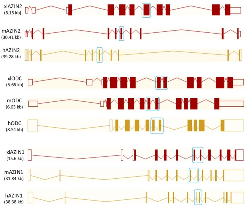
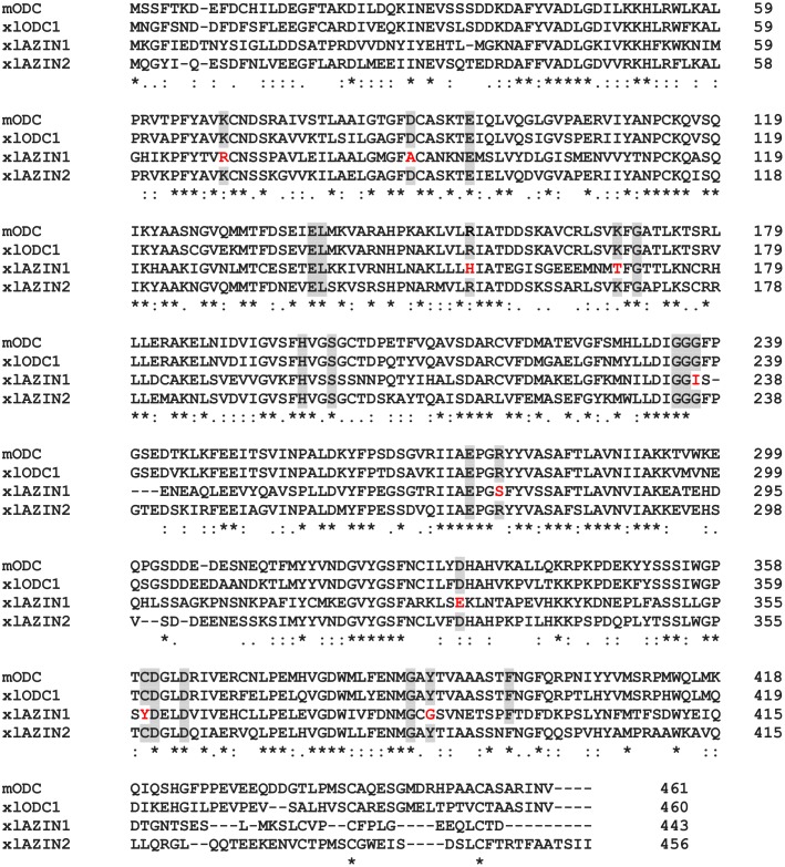
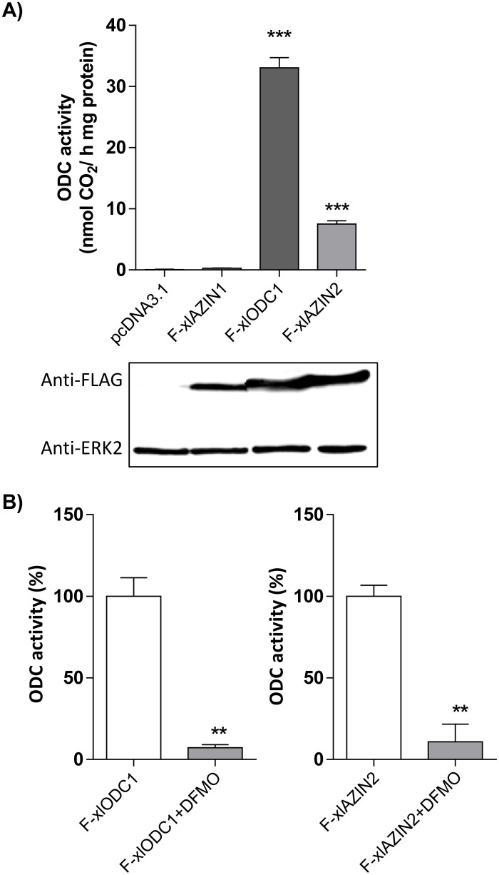
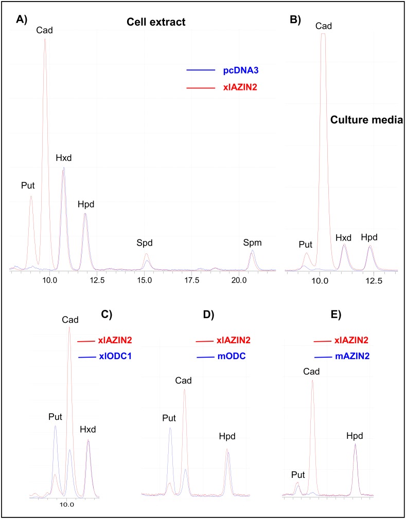
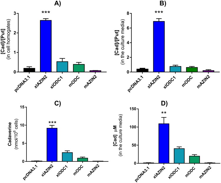
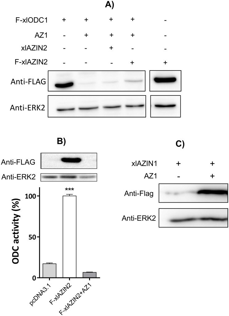
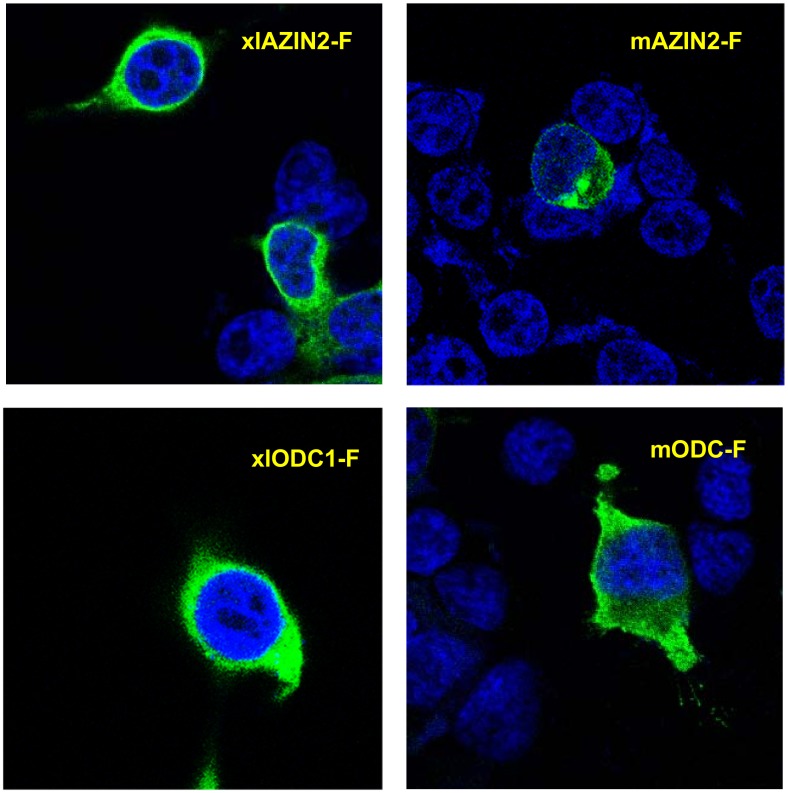
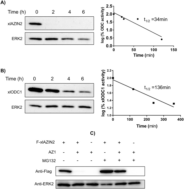
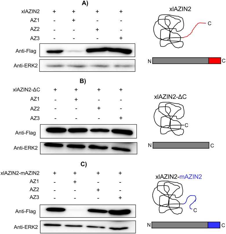
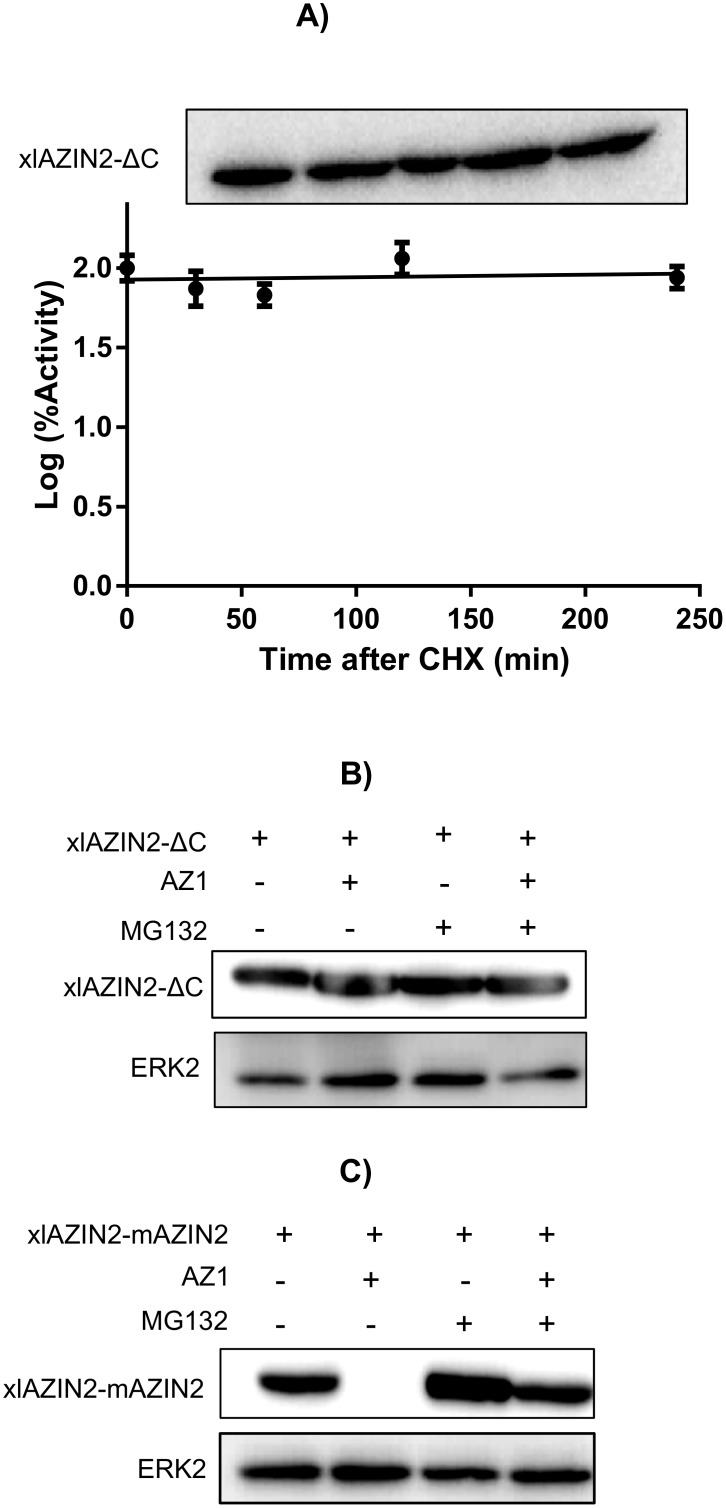
Ornithine decarboxylase (ODC) is a key enzyme in the biosynthesis of polyamines, organic cations that are implicated in many cellular processes. The enzyme is regulated at the post-translational level by an unusual system that includes antizymes (AZs) and antizyme inhibitors (AZINs). Most studies on this complex regulatory mechanism have been focused on human and rodent cells, showing that AZINs (AZIN1 and AZIN2) are homologues of ODC but devoid of enzymatic activity. Little is known about Xenopus ODC and its paralogues, in spite of the relevance of Xenopus as a model organism for biomedical research. We have used the information existing in different genomic databases to compare the functional properties of the amphibian ODC1, AZIN1 and AZIN2/ODC2, by means of transient transfection experiments of HEK293T cells. Whereas the properties of xlODC1 and xlAZIN1 were similar to those reported for their mammalian orthologues, the former catalyzing the decarboxylation of L-ornithine preferentially to that of L-lysine, xlAZIN2/xlODC2 showed important differences with respect to human and mouse AZIN2. xlAZIN2 did not behave as an antizyme inhibitor, but it rather acts as an authentic decarboxylase forming cadaverine, due to its higher affinity to L-lysine than to L-ornithine as substrate; so, in accordance with this, it should be named as lysine decarboxylase (LDC) or lysine/ornithine decarboxylase (LODC). In addition, AZ1 stimulated the degradation of xlAZIN2 by the proteasome, but the removal of the 21 amino acid C-terminal tail, with a sequence quite different to that of mouse or human ODC, made the protein resistant to degradation. Collectively, our results indicate that in Xenopus there is only one antizyme inhibitor (xlAZIN1) and two decarboxylases, xlODC1 and xlLDC, with clear preferences for L-ornithine and L-lysine, respectively.


Conflict of interest statement

The authors have declared that no competing interests exist.

Figures

Fig 1
Fig 1. Genetic structure of mouse and human ODC paralogues, and their comparison with their Xenopus orthologues.
Note that exons 7 and 8 in ODC and AZIN1 are fused in only one exon in AZIN2 (blue boxes). Data obtained from Ensembl (www.ensembl.org).
Fig 2
Fig 2. Comparison of the amino acid sequences of mouse ODC, xlODC1, xlAZIN1 and xlAZIN2 using ClustalW program for multiple sequence alignment.
Asterisks represent amino acid identity; colon and dots represent amino acid similarity between the proteins. Grey background indicates amino acid residues associated with the catalytic activity of mODC that are conserved in the Xenopus laevis homologues. In red: substitutions in these critical residues in xlAZIN1.
Fig 3
Fig 3. Expression of xlODC1, xlAZIN1 and xlAZIN2 in HEK293T transfected cells.
HEK293T cells were transfected with the corresponding constructs of Flag-xlODC1, Flag-xlAZIN1, Flag-xlAZIN2 or empty vector, as indicated in the Experimental Procedures. (A) Top: ODC activity measured in the cell lysates. Bottom: Western blot analysis of the proteins detected using anti-Flag or anti-ERK2 antibodies. Results are expressed as mean±SE, and are representative of three experiments. (***) P<0.001 vs pcDNA3.1 or F-xlODC1. (B) Influence of 1mM alfa-difluoromethylornithine (DFMO) on the ornithine decarboxylase activity of xlODC1 and xlAZIN2 cell lysates. DFMO was added 5h before collecting the cells. (**) P<0.01.
Fig 4
Fig 4. Analysis of the products formed by HEK293T cells transfected with different constructs.
After 16 h of transfection, the culture media was aspirated and the cells collected. An aliquot of the media was concentrated and resuspended in perchloric acid 0.4 M, whereas the cells were homogenized in the same acid (200 μl per well). After centrifugation at 12,000 ×g for 15 min, the supernatants were dansylated and analyzed by HPLC as described in the Experimental section. (A) Overlapped HPLC chromatogram traces of the dansylated extracts from cells transfected with xlAZIN2 (red line) or with the empty vector pcDNA 3.1 (blue line). Hexanediamine (Hxd) and heptanediamine (Hpd) were used as internal standards. Put: putrescine; Cad: cadaverine; Spd: spermidine; Spm: spermine. (B) Overlapped HPLC chromatogram traces corresponding to the dansylated polyamines present in the culture media of cells transfected with xlAZIN2 (red line) or empty vector (blue line). (C) Comparison of the polyamines found in the culture media of cells transfected with xlAZIN2 (red line) with those of xlODC1, mODC and mAZIN2 (blue line).
Fig 5
Fig 5. Cadaverine concentration and Cad/Put ratio in HEK 293T cells transfected with xlAZIN2 or other homologues.
Polyamine levels were analyzed by HPLC in cell homogenates and culture media 16 h after transfection. Cad/Put ratio in cell homogenates (A) or in the culture media (B). Cadaverine concentration in cell homogenates (C) or in the culture media (D). (***) P<0.001 vs the other columns; (**) P<0.01 vs the other columns.
Fig 6
Fig 6. Influence of AZ1 on protein levels of xlODC1 and xlAZIN2.
(A) Western blot of lysates of HEK293T cells co-transfected with xlODC1 and different combinations of AZ1 and xlAZIN2. (B) Western blot and ODC activity of lysates of cells co-transfected with Flag-xlAZIN2 and pcDNA3.1 or AZ1. (***) P<0.001 vs pcDNA3.1 or F-xlAZIN2+AZ1. C) Western blot of lysates of HEK293T cells transfected with xlAZIN1-Flag alone or in combination with AZ1.
Fig 7
Fig 7. Subcellular location of xlODC1 and xlAZIN2 in transfected cells.
Laser scanning confocal micrographs of HEK293T cells transfected with xlODC1, xlAZIN2, mODC or mAZIN2 fused to the Flag epitope. After transfections, cells were fixed, permeabilized and stained with anti-Flag antibody and ALEXA anti-mouse and nuclear DAPI staining, and then examined in a confocal microscope. Flag-proteins are shown in green and nuclei in blue.
Fig 8
Fig 8. Protein stability of xlAZIN2 and xlODC1 in transfected cells.
After 16 h of transfection, either with xlAZIN2 or xlODC1, cells were incubated with 200 μM cycloheximide (CHX), harvested at the indicated times, and lysed in buffer containing a protease inhibitor cocktail. (A) Left: Western blot analysis of xlAZIN2 protein using the anti-Flag antibody; right: decay of ODC activity. (B) Similar experiments with xlODC1. Half-lives of xlAZIN2 and xlODC1 in the transfected cells were calculated by linear regression analysis (GraphPad software). (C) HEK293T cells transfected with xlAZIN2 or xlAZIN2+AZ1were incubated for 5 h with or without the proteasomal inhibitor MG132 (50 μM). xlAZIN2 protein was determined as in (A). ERK2 was used as a loading control.
Fig 9
Fig 9. Influence of the C-terminal region of xlAZIN2 in the degradative process induced by AZs.
HEK293T cells were transfected with: (A) xlAZIN2, (B) xlAZIN2 lacking the 21 C-terminal residues (xlAZIN2-ΔC) or (C) with a construct coding for a chimeric protein with the substitution of the 21 C-terminal residues of xlAZIN2 by the C-terminal segment of mouse AZIN2 (xlAZIN2-mAZIN2). In parallel, each one of the constructs was co-transfected with members of the AZ family (AZ1, AZ2, and AZ3). Western blots were probed with anti-Flag antibody. On the right side, schematic representations of xlAZIN2 and the two mutated proteins.
Fig 10
Fig 10. Protein stability of the mutated forms of xlAZIN2.
(A) After 16 h of transfection with xlAZIN2-ΔC, cells were incubated with 200 μM cycloheximide (CHX), harvested at the indicated times, and lysed in buffer containing a protease inhibitor cocktail. Top: western blot analysis of xlAZIN2-ΔC at different times after CHX addition; bottom: changes in ODC activity after CHX treatment. (B) Influence of the proteasomal inhibitor MG132 (50 μM) on the effect of AZ1 on xlAZIN2-ΔC protein in HEK293T transfected cells. (C) Influence of the proteasomal inhibitor MG132 (50 μM) on the effect of AZ1 on xlAZIN2-mAZIN2 protein in HEK293T transfected cells.

Similar articles

Cited by

References

    1. Pegg AE. Regulation of ornithine decarboxylase. J Biol Chem. 2006; 281(21): 14529–14532. 10.1074/jbc.R500031200 - DOI - PubMed
    1. Cohen S. A Guide to the Polyamines. 1997. Oxford, UK: Oxford University Press.
    1. Igarashi K, Kashiwagi K. Polyamines: Mysterious modulators of cellular functions. Biochem Biophys Res Commun. 2000; 271(3): 559–564. 10.1006/bbrc.2000.2601 - DOI - PubMed
    1. Gerner EW, Meyskens FL. Polyamines and cancer: old molecules, new understanding. Nat Rev Cancer. 2004; 4(10): 781–792. 10.1038/nrc1454 - DOI - PubMed
    1. Igarashi K, Kashiwagi K. Modulation of cellular function by polyamines. Int J Biochem Cell Biol. 2010; 42(1): 39–51. 10.1016/j.biocel.2009.07.009 - DOI - PubMed

Publication types