

Anatomical connections underlying personally-familiar face processing
- PMID: 31509558
- PMCID: PMC6738923
- DOI: 10.1371/journal.pone.0222087
Anatomical connections underlying personally-familiar face processing
Abstract
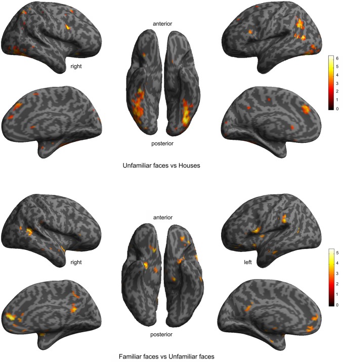
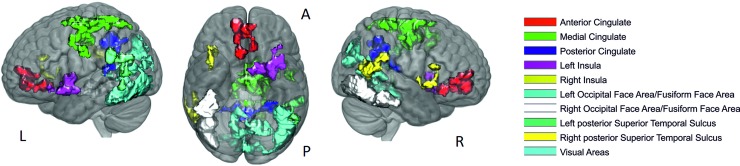
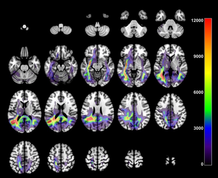
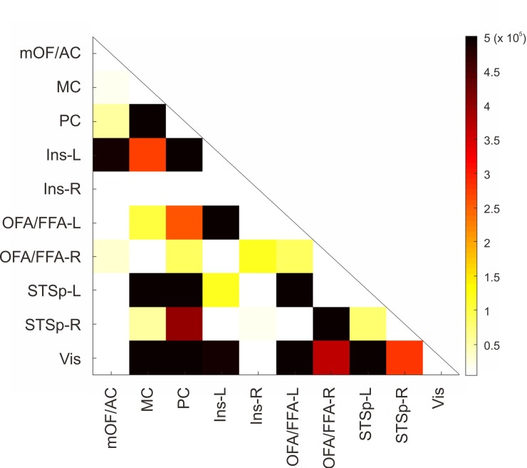
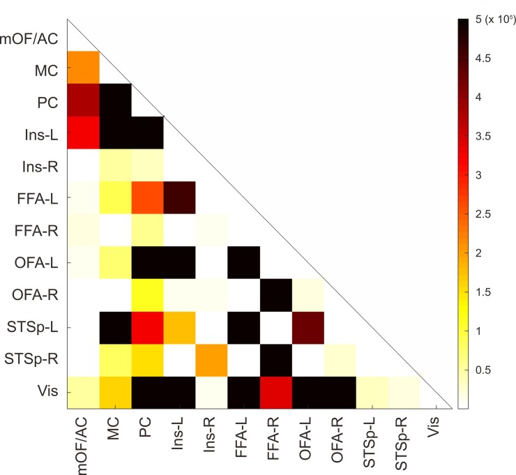
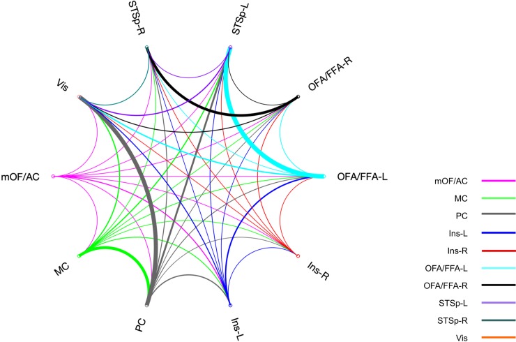
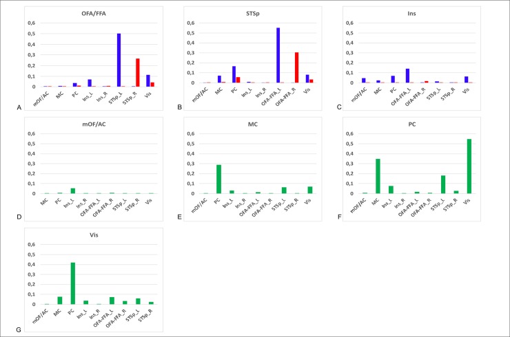
Familiar face processing involves face specific regions (the core face system) as well as other non-specific areas related to processing of person-related information (the extended face system). The connections between core and extended face system areas must be critical for face recognition. Some studies have explored the connectivity pattern of unfamiliar face responding area, but none have explored those areas related to face familiarity processing in the extended system. To study these connections, diffusion weighted imaging with probabilistic tractography was used to estimate the white-matter pathways between core and extended system regions, which were defined from functional magnetic resonance imaging responses to personally-familiar faces. Strong white matter connections were found between occipitotemporal face areas (OFA/FFA) with superior temporal sulcus and insula suggesting the possible existence of direct anatomical connections from face-specific areas to frontal nodes that could underlay the processing of emotional information associated to familiar faces.
Conflict of interest statement
The authors have declared that no competing interests exist.
Figures
Similar articles
-
Direct Structural Connections between Auditory and Visual Motion-Selective Regions in Humans.J Neurosci. 2021 Mar 17;41(11):2393-2405. doi: 10.1523/JNEUROSCI.1552-20.2021. Epub 2021 Jan 29. J Neurosci. 2021. PMID: 33514674 Free PMC article.
-
Effects of face repetition on ventral visual stream connectivity using dynamic causal modelling of fMRI data.Neuroimage. 2022 Dec 1;264:119708. doi: 10.1016/j.neuroimage.2022.119708. Epub 2022 Oct 21. Neuroimage. 2022. PMID: 36280098
-
Two areas for familiar face recognition in the primate brain.Science. 2017 Aug 11;357(6351):591-595. doi: 10.1126/science.aan1139. Science. 2017. PMID: 28798130 Free PMC article.
-
Face Recognition.Curr Neurol Neurosci Rep. 2019 May 30;19(7):41. doi: 10.1007/s11910-019-0960-9. Curr Neurol Neurosci Rep. 2019. PMID: 31144153 Review.
-
The anterior fusiform gyrus: The ghost in the cortical face machine.Neurosci Biobehav Rev. 2024 Mar;158:105535. doi: 10.1016/j.neubiorev.2024.105535. Epub 2024 Jan 6. Neurosci Biobehav Rev. 2024. PMID: 38191080 Review.
Cited by
-
Spatio-temporal brain dynamics of self-identity: an EEG source analysis of the current and past self.Brain Struct Funct. 2022 Jul;227(6):2167-2179. doi: 10.1007/s00429-022-02515-9. Epub 2022 Jun 7. Brain Struct Funct. 2022. PMID: 35672533 Free PMC article.
-
Application of Empirical Mode Decomposition for Decoding Perception of Faces Using Magnetoencephalography.Sensors (Basel). 2021 Sep 17;21(18):6235. doi: 10.3390/s21186235. Sensors (Basel). 2021. PMID: 34577441 Free PMC article.
References
Publication types
MeSH terms
Associated data
Grants and funding
LinkOut - more resources
Full Text Sources
