. 2019 Sep 10;28(11):2878-2891.e5.
doi: 10.1016/j.celrep.2019.08.024.

Orphan Nuclear Receptor NR2F6 Suppresses T Follicular Helper Cell Accumulation through Regulation of IL-21

Affiliations

Orphan Nuclear Receptor NR2F6 Suppresses T Follicular Helper Cell Accumulation through Regulation of IL-21

William J Olson et al. Cell Rep. .

Abstract

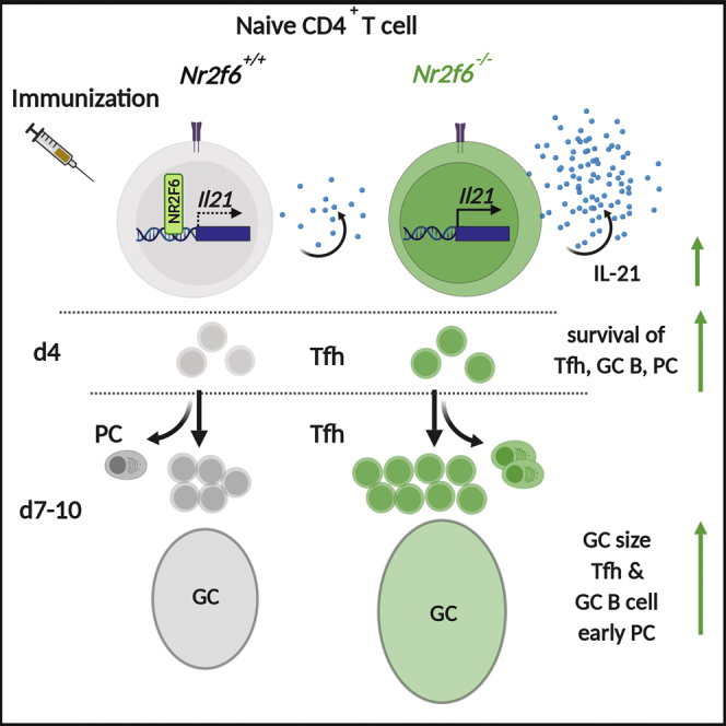
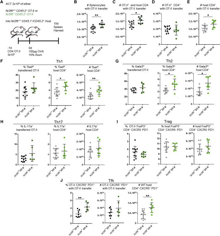
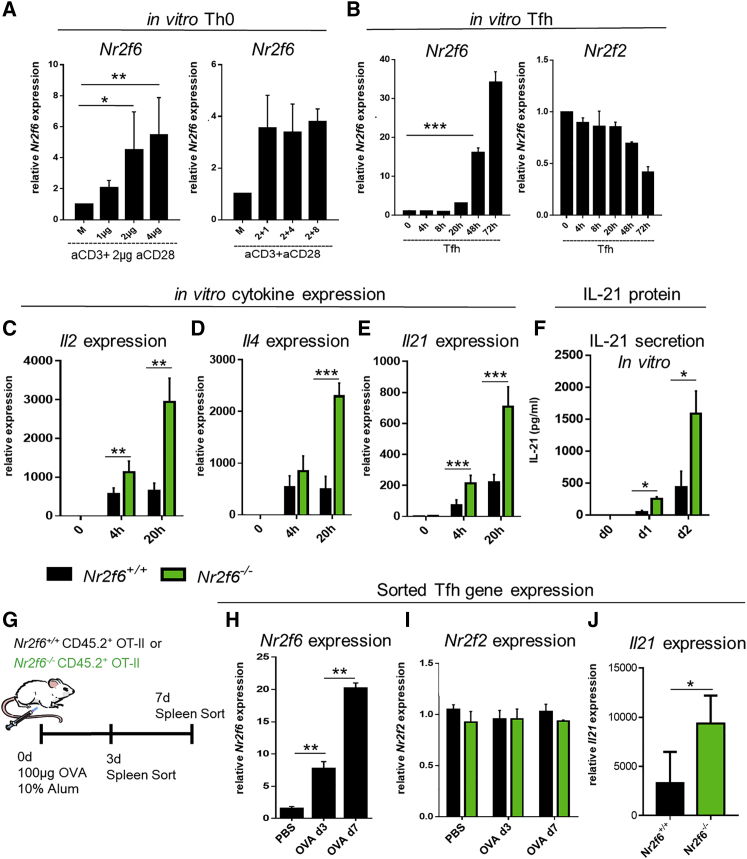
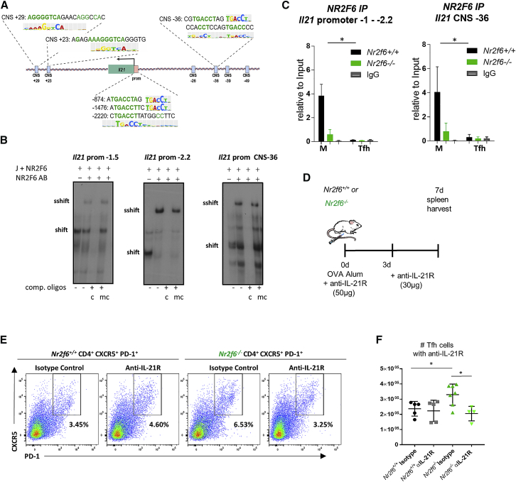
CD4 T follicular helper (Tfh) cells are specialized in helping B cells during the germinal center (GC) reaction and ultimately promote long-term humoral immunity. Here we report that loss of the nuclear orphan receptor NR2F6 causes enhanced survival and accumulation of Tfh cells, GC B cells, and plasma cells (PCs) following T cell-dependent immunization. Nr2f6-deficient CD4 T cell dysfunction is the primary cause of cell accumulation. Cytokine expression in Nr2f6-deficient Tfh cells is dysregulated, and Il21 expression is enhanced. Mechanistically, NR2F6 binds directly to the interleukin 21 (IL-21) promoter and a conserved noncoding sequence (CNS) near the Il21 gene in resting CD4+ T cells. During Tfh cell differentiation, this direct NR2F6 DNA interaction is abolished. Enhanced Tfh cell accumulation in Nr2f6-deficient mice can be reverted by blocking IL-21R signaling. Thus, NR2F6 is a critical negative regulator of IL-21 cytokine production in Tfh cells and prevents excessive Tfh cell accumulation.

Keywords: IL-21; T follicular helper cell; autoimmunity; germinal center reaction; immunization; nuclear receptor; systemic lupus erythematosus.


Conflict of interest statement

The authors declare no competing interests.

Figures

None
Graphical abstract
Figure 1
Figure 1
Nr2f6 Loss Leads to Tfh Cell Accumulation and Increased GC Responses following OVA-Alum Immunization (A) Experimental setup used for OVA-alum immunization of Nr2f6+/+ or Nr2f6−/− mice. (B and C) Total counts of Nr2f6+/+ or Nr2f6−/− splenocytes, B220+ cells, and CD4+ cells on (B) day 4 or (C) day 10 after immunization. (D and E) Splenic Tfh cell numbers on (D) day 4 or (E) day 10 after immunization; representative flow cytometry plots for Nr2f6+/+ and Nr2f6−/− Tfh cells, total Tfh cell counts, and Tfh cell frequency within CD4 cells. Total Tfh cell counts are shown as total live non-Tfr cells (annexin V, FoxP3, and 7-AAD). (F) Total germinal center B cell counts on day 4. (G) Day 10 representative flow cytometry plots, with total cell counts. (H) GC LZ and DZ distribution in Nr2f6+/+ or Nr2f6−/− GCs 10 days after immunization. (I and J) PC counts on day 4 (I), and representative flow cytometry plots and cell counts on day 10 (J). (K) Immunofluorescence staining for CD3 (white), B220 (red), and peanut agglutinin (PNA) (green) on 5 μM sections of spleens harvested day 10 after OVA immunization. (L and M) Frequency of live Tfh cells, GC B cells, and PCs (annexin V 7-AAD) on (L) day 4 or (M) day 10 after OVA-alum immunization. (N) Tfr (CD4+, CXCR5+, PD-1+, and FoxP3+) total cell counts on day 4 (left panel), and the Tfh cell to Tfr cell ratio (right panel). (O) Representative FoxP3 staining of Nr2f6+/+ or Nr2f6−/− CD4+ CXCR5+ PD-1+ T cells, total Tfr cell counts, total Tfr frequency of all CD4 cells, and the Tfh cell to Tfr cell ratio on day 10 post-immunization. Data shown are from at least two separate experiments with n ≥ 5. The middle bar represents the average for the dataset. Error bars represent SD, and an asterisk indicates statistically significant differences calculated using two-tailed Student’s t test or Mann-Whitney U test. A p value of <0.05 was considered statistically significant. p < 0.05; ∗∗p < 0.01; ∗∗∗p < 0.001.
Figure 2
Figure 2
Nr2f6 Deficiency Does Not Alter Affinity Maturation but Affects Antigen-Specific Memory B Cells (A) Nr2f6+/+ or Nr2f6−/− mice were immunized with NP-CGG in alum; relative binding of high (18) or low (1.7) ratio haptenated NP-BSA-coated plates was measured by ELISA. IgG1 relative affinity is shown from serum collected 10, 20, or 30 days after immunization. High-affinity (NP1.7) or total IgG1 binding (NP18) titers from one representative ELISA experiment are shown. (B) Sera was collected, and ELISA was performed for indicated antibodies 20 days after immunization, optical density 50 (OD50) is shown. (C) Experimental scheme. (D) Nr2f6+/+ or Nr2f6−/− mice were immunized with NP-CGG, and spleens were harvested on day 90; antigen-specific memory B cells were stained with NP-PE and CD19. Representative flow cytometry plots are shown in the left two panels, frequency and total cell count of this population are shown in the right two panels. (E–G) CD80 and PD-L2 were investigated by flow cytometry on IgG1 (E) or IgM (F) antigen-specific memory B cells from both Nr2f6+/+ or Nr2f6−/− genotypes. Recall responses were tested as described in the scheme (G). (H) Total NP-specific, IgD, and dump gate negative (Gr-1, F4/80, CD4, and CD8) cells, proliferation, and dead cell frequency were investigated by Ki67 and efluor450 fixable dye staining. (I and J) GC (I) and PC (J) responses from this population were investigated by flow cytometry. Representative flow cytometry plots and total counts are shown. Memory results are representative of two independent experiments with n ≥ 5. The middle bar represents the dataset average. Error bars represent SD, and an asterisk indicates statistically significant differences calculated using two-tailed Student’s t test. A p value of <0.05 was considered statistically significant. p < 0.05; ∗∗p < 0.01; ∗∗∗p < 0.001.
Figure 3
Figure 3
Nr2f6-Deficient B Cells Do Not Contribute to the GC Phenotype of Nr2f6−/− Mice (A) Experimental scheme showing CD4 depletion of Nr2f6+/+ or Nr2f6−/− host mice followed by Nr2f6+/+ OT-II transfer. (B) Splenocyte stain from wild-type control mouse, with no depletion or OT-II transfer, harvested 7 days after OVA-alum immunization and stained for CD4, Vα2, and Vβ5. (C) Representative plots of Vα2+ Vβ5+ CD4+ T cells 7 days after immunization and 8 days after OT-II transfer. Combined frequency and total cell counts are displayed in the right two panels. (D) Vα2 Vβ5 CD4+ CXCR5+ PD-1+ T cell (host) frequency and total cell counts are displayed with representative flow cytometry plots. (E) Frequency of the Tfh cell population from transferred OT-II cells (CD4+ Vα2+ Vβ5+) and total cell counts are displayed with representative flow cytometry plots. (F and G) Host germinal center B cell (F) and plasma cell frequency (G), as well as total cell counts, are displayed. Data shown are from two independent experiments with n ≥ 6. The middle bar represents the dataset average. Error bars represent SD, and an asterisk indicates statistically significant differences calculated using Student’s t test. A p value of <0.05 was considered statistically significant. p < 0.05; ∗∗p < 0.01; ∗∗∗p < 0.001.
Figure 4
Figure 4
Adoptive Cell Transfer of Nr2f6-Deficient OT-II T Cells into Wild-Type Hosts Results in Increased Host CD4 Tfh, Th1, and Th2 Subsets (A) Experimental scheme showing adoptive transfer of 3 × 106 OT-II CD4+CD45.2+Nr2f6+/+ (black) or Nr2f6−/− (green) into Nr2f6+/+ CD45.1/CD45.2 congenic mice with OVA-alum immunization (n ≥ 7 per group). (B) Total splenocytes, with Nr2f6+/+ or Nr2f6−/− OT-II adoptive cell transfer. (C) Total CD4 T cell counts, including both host and transferred OT-II cells. (D and E) Transferred OT-II T cell counts (D) and total host CD4 T cell counts (E). (F) Frequency of Th1 cells from the indicated OT-II populations or host cells (left and middle panels). Total host Th1 cells in the spleen with indicated OT-II T cell transfer (right panel). (G) Th2 as a frequency of OT-II or host CD4 T cell populations and total Th2 cells from the host CD4 T cells. (H) Frequency and total cell count of Th17 cells. (I) Treg subset defined as CXCR5 PD-1 CD4+ FoxP3+ from both OT-II transferred cells and host cells. (J) Tfh cells defined as CXCR5+ PD-1+ Foxp3 CD4+ from OT-II and host cells. Results shown are derived from three independent experiments (n ≥ 7). The middle bar shows an average of each dataset. Error bars represent SD, and an asterisk indicates statistically significant differences between genotypes calculated using Student’s t test or Mann-Whitney U test. A p value of <0.05 was considered statistically significant. p < 0.05; ∗∗p < 0.01.
Figure 5
Figure 5
NR2F6 Suppresses Il21 Expression in Tfh Cells In Vitro and In Vivo (A) Naive Nr2f6+/+ CD4+ T cells were stimulated for 24 h with indicated amounts of anti-CD3 and a fixed amount (2 μg/mL) of anti-CD28 (left panel). Alternatively, anti-CD3 was kept constant, and anti-CD28 was added at the indicated dose (right panel). qRT-PCR performed for Nr2f6 expression is displayed; values are shown relative to gapdh expression. (B) Wild-type CD4 T cells were cultured under Tfh cell-polarizing conditions, and qRT-PCR was used to determine Nr2f6 expression at the indicated time points. Expression of the closely related nuclear receptor Nr2f2 was also determined under these conditions (right panel). (C–E) Cytokine expression of (C) Il2, (D) Il4, and (E) Il21 was determined in in vitro Tfh cell culture by qRT-PCR at the indicated time points. (F) IL-21 secretion into culture media was measured using Bioplex technology. (G) Scheme for Nr2f6+/+ OT-II and Nr2f6−/− OT-II mouse immunization and Tfh cell sorting. (H) Day 3 and day 7 Nr2f6 expression from sorted Nr2f6+/+ OT-II Tfh cells. (I) Nr2f2 expression from Nr2f6+/+ OT-II and Nr2f6−/− OT-II Tfh cells sorted as in (H). (J) Il21 expression from day 7 sorted Nr2f6+/+ OT-II or Nr2f6−/− OT-II Tfh cells. Data shown are from two independent experiments with n ≥ 4. The middle bar represents the dataset average. The data are presented as the percentage of input samples before immunoprecipitation. Error bars represent SD, and an asterisk indicates statistically significant differences between genotypes calculated using Student’s t test. A p value of <0.05 was considered statistically significant. p < 0.05; ∗∗p < 0.01; ∗∗∗p < 0.001.
Figure 6
Figure 6
NR2F6 Directly Binds to the Il21 Promoter and CNS −36 in Resting CD4 T Cells, and Inhibition of IL-21 Signaling Reduces Tfh Cell Accumulation (A) Putative NR2F6 binding sites of the mouse Il21 promoter and CNS regions are shown. (B) NR2F6 binding to the Il21 promoter at −1.5 kb, −2.2 kb, or CNS −36 by EMSA nuclear extracts from Jurkat cells (J+ NR2F6) transfected with pEFneo Nr2f6 plasmid. To validate binding specificity, consensus (c) or mutated (mc) oligonucleotides were added in excess as unlabeled competition oligonucleotides. Where indicated, an anti-NR2F6 antibody was added. One representative experiment out of three is shown. (C) NR2F6 binding to the Il21 promoter at −1 to −2.2 kb or CNS −36 was investigated by ChIP. Nr2f6+/+ or Nr2f6–/– CD4 cells either resting or activated under Tfh-differentiating conditions were used with anti-NR2F6 or IgG control precipitation, Il21 promoter or CNS sequence was quantified by qPCR. Data are presented as the percentage of input. Differences between genotypes were calculated using log-transformed data following a linear mixed-effects model fit by restricted maximum likelihood (REML) and was analyzed via ANOVA; interaction plots are shown in Figures S6F–S6H. (D) Scheme for anti-IL-21R block within OVA-alum-immunized Nr2f6+/+ or Nr2f6−/− mice. Wild-type or Nr2f6−/− mice were treated with anti-IL-21R antibody or isotype control at the same time as OVA immunization (50 μg) and 3 days later (30 μg). (E and F) Representative Tfh cell flow cytometry data (E) and total Tfh cell numbers (F) are shown. Data shown are from two independent experiments with n ≥ 4. Error bars represent SD, and an asterisk indicates statistically significant differences between genotypes calculated using Student’s t test, Mann-Whitney U test, or ANOVA. A p value of <0.05 was considered statistically significant. p < 0.05; ∗∗p < 0.01; ∗∗∗p < 0.001.

Similar articles

Cited by

References

    1. Aprahamian T.R., Bonegio R.G., Weitzner Z., Gharakhanian R., Rifkin I.R. Peroxisome proliferator-activated receptor gamma agonists in the prevention and treatment of murine systemic lupus erythematosus. Immunology. 2014;142:363–373. - PMC - PubMed
    1. Baumjohann D., Preite S., Reboldi A., Ronchi F., Ansel K.M., Lanzavecchia A., Sallusto F. Persistent antigen and germinal center B cells sustain T follicular helper cell responses and phenotype. Immunity. 2013;38:596–605. - PubMed
    1. Bell J., Gray D. Antigen-capturing cells can masquerade as memory B cells. J. Exp. Med. 2003;197:1233–1244. - PMC - PubMed
    1. Bubier J.A., Sproule T.J., Foreman O., Spolski R., Shaffer D.J., Morse H.C., 3rd, Leonard W.J., Roopenian D.C. A critical role for IL-21 receptor signaling in the pathogenesis of systemic lupus erythematosus in BXSB-Yaa mice. Proc. Natl. Acad. Sci. USA. 2009;106:1518–1523. - PMC - PubMed
    1. Burris T.P., Solt L.A., Wang Y., Crumbley C., Banerjee S., Griffett K., Lundasen T., Hughes T., Kojetin D.J. Nuclear receptors and their selective pharmacologic modulators. Pharmacol. Rev. 2013;65:710–778. - PMC - PubMed

MeSH terms