NAD hydrolysis by the tuberculosis necrotizing toxin induces lethal oxidative stress in macrophages
- PMID: 31509891
- PMCID: PMC6925628
- DOI: 10.1111/cmi.13115
NAD hydrolysis by the tuberculosis necrotizing toxin induces lethal oxidative stress in macrophages
Abstract
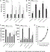
Mycobacterium tuberculosis (Mtb) kills infected macrophages through necroptosis, a programmed cell death that enhances mycobacterial replication and dissemination. The tuberculosis necrotizing toxin (TNT) is the major cytotoxicity factor of Mtb in macrophages and induces necroptosis by NAD+ hydrolysis. Here, we show that the catalytic activity of TNT triggers the production of reactive oxygen species (ROS) in Mtb-infected macrophages causing cell death and promoting mycobacterial replication. TNT induces ROS formation both by activating necroptosis and by a necroptosis-independent mechanism. Most of the detected ROS originate in mitochondria as a consequence of opening the mitochondrial permeability transition pore. However, a significant part of ROS is produced by mechanisms independent of TNT and necroptosis. Expressing only the tnt gene in Jurkat T-cells also induces lethal ROS formation indicating that these molecular mechanisms are not restricted to macrophages. Both the antioxidant N-acetyl-cysteine and replenishment of NAD+ by providing nicotinamide reduce ROS levels in Mtb-infected macrophages, protect them from cell death, and restrict mycobacterial replication. Our results indicate that a host-directed therapy combining replenishment of NAD+ with inhibition of necroptosis and/or antioxidants might improve the health status of TB patients and augment antibacterial TB chemotherapy.
Keywords: TNT; macrophages; necroptosis; nicotinamide adenine dinucleotide; reactive oxygen species.
© 2019 John Wiley & Sons Ltd.
Conflict of interest statement
Competing financial interests: The authors declare no financial conflict of interest.
Figures


Similar articles
-
NAD+ Depletion Triggers Macrophage Necroptosis, a Cell Death Pathway Exploited by Mycobacterium tuberculosis.Cell Rep. 2018 Jul 10;24(2):429-440. doi: 10.1016/j.celrep.2018.06.042. Cell Rep. 2018. PMID: 29996103 Free PMC article.
-
The tuberculosis necrotizing toxin kills macrophages by hydrolyzing NAD.Nat Struct Mol Biol. 2015 Sep;22(9):672-8. doi: 10.1038/nsmb.3064. Epub 2015 Aug 3. Nat Struct Mol Biol. 2015. PMID: 26237511 Free PMC article.
-
The tuberculosis necrotizing toxin is an NAD+ and NADP+ glycohydrolase with distinct enzymatic properties.J Biol Chem. 2019 Mar 1;294(9):3024-3036. doi: 10.1074/jbc.RA118.005832. Epub 2018 Dec 28. J Biol Chem. 2019. PMID: 30593509 Free PMC article.
-
Cell death at the cross roads of host-pathogen interaction in Mycobacterium tuberculosis infection.Tuberculosis (Edinb). 2018 Dec;113:99-121. doi: 10.1016/j.tube.2018.09.007. Epub 2018 Sep 29. Tuberculosis (Edinb). 2018. PMID: 30514519 Review.
-
ADP-ribosylation of membrane proteins: unveiling the secrets of a crucial regulatory mechanism in mammalian cells.Ann Med. 2006;38(3):188-99. doi: 10.1080/07853890600655499. Ann Med. 2006. PMID: 16720433 Review.
Cited by
-
The cause-effect relation of tuberculosis on incidence of diabetes mellitus.Front Cell Infect Microbiol. 2023 Jun 26;13:1134036. doi: 10.3389/fcimb.2023.1134036. eCollection 2023. Front Cell Infect Microbiol. 2023. PMID: 37434784 Free PMC article. Review.
-
Mycobacterial Control of Host Mitochondria: Bioenergetic and Metabolic Changes Shaping Cell Fate and Infection Outcome.Front Cell Infect Microbiol. 2020 Sep 30;10:457. doi: 10.3389/fcimb.2020.00457. eCollection 2020. Front Cell Infect Microbiol. 2020. PMID: 33102245 Free PMC article. Review.
-
NAD+ Metabolism, Metabolic Stress, and Infection.Front Mol Biosci. 2021 May 19;8:686412. doi: 10.3389/fmolb.2021.686412. eCollection 2021. Front Mol Biosci. 2021. PMID: 34095234 Free PMC article. Review.
-
Untargeted metabolomics analysis reveals Mycobacterium tuberculosis strain H37Rv specifically induces tryptophan metabolism in human macrophages.BMC Microbiol. 2022 Oct 17;22(1):249. doi: 10.1186/s12866-022-02659-y. BMC Microbiol. 2022. PMID: 36253713 Free PMC article.
-
Adjunct N-Acetylcysteine Treatment in Hospitalized Patients With HIV-Associated Tuberculosis Dampens the Oxidative Stress in Peripheral Blood: Results From the RIPENACTB Study Trial.Front Immunol. 2021 Feb 4;11:602589. doi: 10.3389/fimmu.2020.602589. eCollection 2020. Front Immunol. 2021. PMID: 33613521 Free PMC article. Clinical Trial.
References
-
- Awodele O, Olayemi SO, Nwite JA, & Adeyemo TA (2012). Investigation of the levels of oxidative stress parameters in HIV and HIV-TB co-infected patients. J Infect Dev Ctries, 6(1), 79–85. - PubMed
Publication types
MeSH terms
Substances
Grants and funding
LinkOut - more resources
Full Text Sources
Molecular Biology Databases

