Review
. 2019 Sep 10;8(9):1057.
doi: 10.3390/cells8091057.

The Molecular Links between Cell Death and Inflammasome

Affiliations
Review

The Molecular Links between Cell Death and Inflammasome

Kwang-Ho Lee et al. Cells. .

Abstract

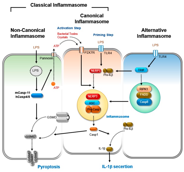
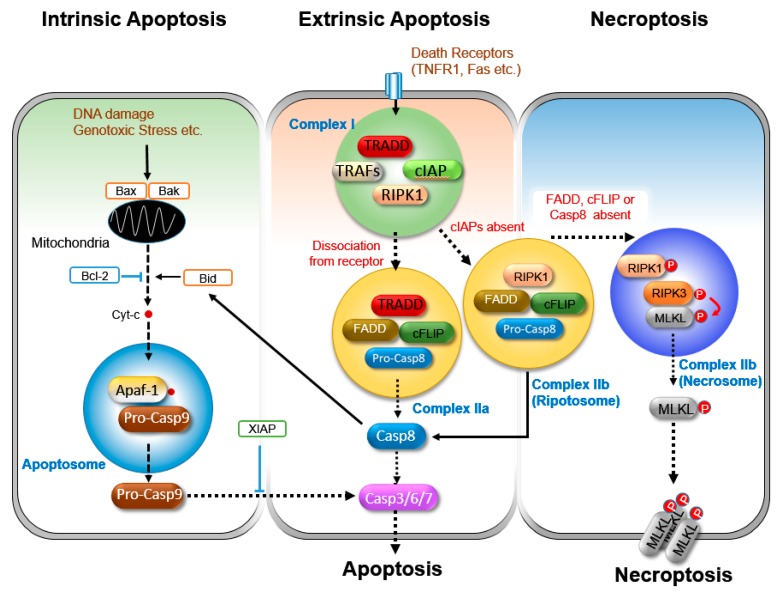
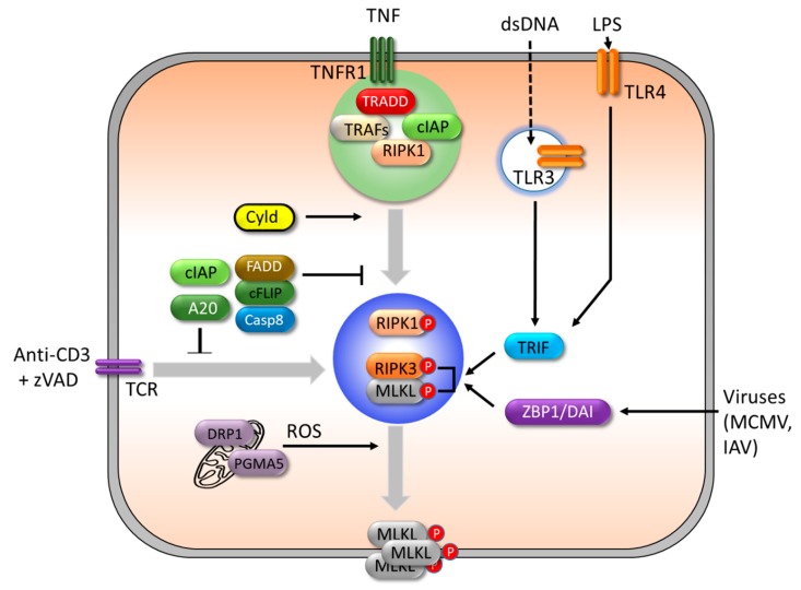
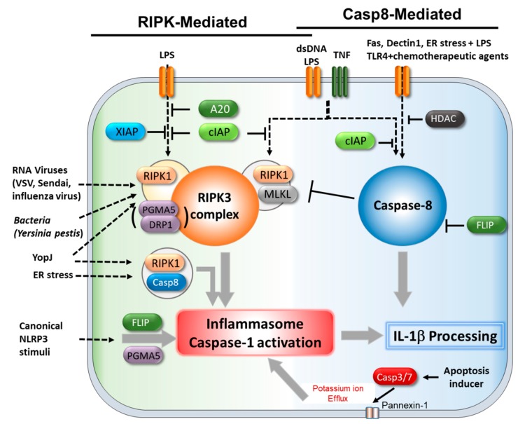
Programmed cell death pathways and inflammasome activation pathways can be genetically and functionally separated. Inflammasomes are specialized protein complexes that process pro-inflammatory cytokines, interleukin-1β (IL-1β), and IL-18 to bioactive forms for protection from a wide range of pathogens, as well as environmental and host-derived danger molecules. Programmed cell death has been extensively studied, and its role in the development, homeostasis, and control of infection and danger is widely appreciated. Apoptosis and the recently recognized necroptosis are the best-characterized forms of programmed death, and the interplay between them through death receptor signaling is also being studied. Moreover, growing evidence suggests that many of the signaling molecules known to regulate programmed cell death can also modulate inflammasome activation in a cell-intrinsic manner. Therefore, in this review, we will discuss the current knowledge concerning the role of the signaling molecules originally associated with programmed cell death in the activation of inflammasome and IL-1β processing.

Keywords: Caspase-8; DRP1; MLKL; PGAM5; RIPK1/3; apoptosis; inflammasome; necroptosis; programmed cell death.


Conflict of interest statement

The authors declare no conflict of interest.

Figures

Figure 1
Figure 1
A simple depiction of signaling pathways for the activation of classical NLRP3 inflammasome (canonical and non-canonical) and alternative inflammasome. Canonical inflammasome activation includes a priming and activation step. The priming step, by pathogen-associated molecular patterns (PAMPs) and cytokines, leads to the expression of inflammasome components, NLRP3, and pro-IL-1β through the NF-κB pathway. The activation step is triggered by damage-associated molecular patterns (DAMPs), such as ATP, crystals, pore-forming toxins, and metabolites, which induce the formation of the NLRP3 inflammasome complex, where caspase-1 is activated. Activated caspase-1 in turn cleaves pro-IL-1β for the production and secretion of active IL-1β. It also cleaves gasdermin D (GSMD), which leads to the formation of membrane pores and triggers pyroptosis. Non-canonical NLRP3-inflammasome is initiated by sensing cytosolic LPS by caspase-11/-4/-5. Then, activated caspase-11/-5/-4 cleaves GSDMD, which leads to pyroptosis. In parallel, the activated caspase-11/-4/-5 activates pannexin-1, leading to ATP release and K+ efflux in order to derive NLRP3 inflammasome activation and IL-1β secretion. In contrast to classical inflammasome activation, alternative inflammasome activation can be triggered by a single signal. LPS stimulation induces the activation of NLRP3 inflammasome via a TLR4-TRIF-RIPK1-FADD-CASP8 signaling axis, independently of the potassium efflux. The alternative inflammasome activation does not induce pyroptosis.
Figure 2
Figure 2
A simple depiction of the pathways for intrinsic and extrinsic apoptosis and necroptosis. The triggering of intrinsic apoptosis induces the mitochondrial membrane potential disruption and the release of cytochrome C into the cytoplasm. Then, cytochrome C interacts with apoptotic protease activating factor 1 (APAF1) and pro-caspase-9 to form a complex, named the apoptosome, where pro-caspase 9 is activated by the cleavage. The activated caspase-9 then activates the executioner caspases-3, -6, and -7 to execute cell death. This pathway is promoted by pro-apoptotic members of the Bcl2 family, such as BAK, BAX, and BID, activated by caspase-8, and is suppressed by anti-apoptotic proteins BCL-2, BCl-XL, and XIAP. The extrinsic apoptosis pathway is initiated by the activation of the death receptors of the TNF super family. Upon TNF stimulation, a receptor bound complex (complex I) is formed by RIPK1, TRADD, cIAP, and TRAFs, which essentially prevents the transition to the cell death pathway. However, TRADD/RIPK1 dissociation from the receptor, or TNF stimulation in the absence of cIAPs activity, causes the formation of different complexes, called complex IIa or complex IIb (ripoptosome), by interacting with FADD and caspase-8, where caspase-8 is activated. Then, active caspase-8 induces apoptosis through the activation of effector caspase-3/-6/-7, or by the cleavage of BID to promote cytochrome C release. TNF stimulation under the inhibition of both caspase-8 activity and cIAPs leads to the formation of another complex IIb (necrosome), where RIPK1, RIPK3, and MLKL are activated through phosphorylation. Then, activated MLKL forms an oligomer and translocates to the plasma membrane to execute necroptosis.
Figure 3
Figure 3
A simple depiction of the triggers and regulators of necroptosis. Upon TNF stimulation, cIAPs in the complex I inhibit necroptosis and execute cell survival signaling. Upon activation of TLRs by dsDNA, LPS, or viral infection, RIPK1-independent and RIPK3-mediated necroptosis is triggered by two RHIM-containing adaptor proteins, TRIF and ZBP1/DAI. T cell receptor triggering with caspase inhibitor (zVAD) also induces RIPK3-dependent necroptosis. The deubiquitination of RIPK1 by CYLD facilitates the formation of necrosome and promotes necroptosis. Another deubiquitinase, A20, also suppresses necroptosis through deubiquitinating RIPK1 or RIPK3. Phosphoglycerate mutase family member 5 (PGAM5) and Dynamin related protein 1 (Drp1) induce reactive oxygen species (ROS) production in the mitochondria and contribute to plasma membrane rupture.
Figure 4
Figure 4
Roles of necroptosis molecules in inflammasome activation and IL-1β processing. Apoptosis- or necroptosis-related molecules contribute to inflammasome activation and IL-1β processing through two distinctive pathways. One is through the protein kinase RIPK-complex, the other one is through caspase-8. Caspase-8 can directly cleaves pro-IL-1β upon stimulation with stimuli such as FasL, Dectin-1, and LPS combined with chemotherapeutic drugs, as well as the inhibition of HDAC. The caspase-8 function in these processes could be inhibited by c-FLIP. Caspase-8 can also promote the activation of inflammasome or caspase-1 in response to YopJ and endoplasmic reticulum (ER) stress. The disruption of some genes, including A20, cIAPs, and XIAP, induces RIPK3-complex-mediated inflammasome activation upon stimulation with a TLR-agonist and TNF. Pathogenic infection, such as RNA viruses and bacteria, induce inflammasome activation through the RIPK1–RIPK3 complex. Depending on the stimuli, the RIPK1–RIPK3 complex promotes inflammasome activation with the help of DRP1 and PGAM5. PGAM5 and c-FLIP contribute to canonical NLRP3 inflammasome activation induced by LPS and ATP or monosodium urate (MSU). The disruption of caspase-8 leads to MLKL–RIPK3-mediated inflammasome activation. A caspase-3/-7-mediated pannexin-1activation during apoptosis promotes NLRP3 inflammasome via potassium efflux.

Similar articles

Cited by

References

    1. Dinarello C.A. Immunological and inflammatory functions of the interleukin-1 family. Annu. Rev. Immunol. 2009;27:519–550. doi: 10.1146/annurev.immunol.021908.132612. - DOI - PubMed
    1. Lamkanfi M., Dixit V.M. Mechanisms and functions of inflammasomes. Cell. 2014;157:1013–1022. doi: 10.1016/j.cell.2014.04.007. - DOI - PubMed
    1. He Y., Hara H., Nunez G. Mechanism and Regulation of NLRP3 Inflammasome Activation. Trends Biochem. Sci. 2016;41:1012–1021. doi: 10.1016/j.tibs.2016.09.002. - DOI - PMC - PubMed
    1. Cordoba-Rodriguez R., Fang H., Lankford C.S., Frucht D.M. Anthrax lethal toxin rapidly activates caspase-1/ICE and induces extracellular release of interleukin (IL)-1beta and IL-18. J. Biol. Chem. 2004;279:20563–20566. doi: 10.1074/jbc.C300539200. - DOI - PubMed
    1. Cirelli K.M., Gorfu G., Hassan M.A., Printz M., Crown D., Leppla S.H., Grigg M.E., Saeij J.P., Moayeri M. Inflammasome sensor NLRP1 controls rat macrophage susceptibility to Toxoplasma gondii. PLoS Pathog. 2014;10:e1003927. doi: 10.1371/journal.ppat.1003927. - DOI - PMC - PubMed

Publication types

MeSH terms

LinkOut - more resources