

Perivascular Adipocytes in Vascular Disease
- PMID: 31510794
- PMCID: PMC6812617
- DOI: 10.1161/ATVBAHA.119.312304
Perivascular Adipocytes in Vascular Disease
Abstract
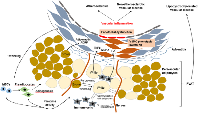
Perivascular adipocytes residing in the vascular adventitia are recognized as distinct endocrine cells capable of responding to inflammatory stimuli and communicating with the sympathetic nervous system and adjacent blood vessel cells, thereby releasing adipocytokines and other signaling mediators to maintain vascular homeostasis. Perivascular adipocytes exhibit phenotypic heterogeneity (both white and brown adipocytes) and become dysfunctional in conditions, such as diet-induced obesity, thus promoting vascular inflammation, vasoconstriction, and smooth muscle cell proliferation to potentially contribute to the development of vascular diseases, such as atherosclerosis, hypertension, and aortic aneurysms. Although accumulating data have advanced our understanding of the role of perivascular adipocytes in modulating vascular function, their impact on vascular disease, particularly in humans, remains to be fully defined. This brief review will discuss the mechanisms whereby perivascular adipocytes regulate vascular disease, with a particular emphasis on recent findings and current limitations in the field of research.
Keywords: adipocyte; adipokine; inflammation; obesity; vasculopathy.
Figures
References
-
- Chatterjee TK, Aronow BJ, Tong WS, Manka D, Tang Y, Bogdanov VY, Unruh D, Blomkalns AL, Piegore MG Jr, Weintraub DS, Rudich SM, Kuhel DG, Hui DY, Weintraub NL. Human coronary artery perivascular adipocytes overexpress genes responsible for regulating vascular morphology, inflammation, and hemostasis. Physiol Genomics. 2013;45:697–709. - PMC - PubMed
Publication types
MeSH terms
Substances
Grants and funding
LinkOut - more resources
Full Text Sources
Medical
