

The differences of gonadal hormones and uterine transcriptome during shell calcification of hens laying hard or weak-shelled eggs
- PMID: 31510913
- PMCID: PMC6737649
- DOI: 10.1186/s12864-019-6017-2
The differences of gonadal hormones and uterine transcriptome during shell calcification of hens laying hard or weak-shelled eggs
Abstract
Background: Eggshell breaking strength is critical to reduce egg breaking rate and avoid economic loss. The process of eggshell calcification initiates with the egg entering the uterus and lasts about 18 h. It follows a temporal sequence corresponding to the initiation, growth and termination periods of shell calcification. During each period of shell calcification, our study investigated the differences of gonadal hormones and uterine transcriptome in laying hens producing a high or low breaking strength shell.
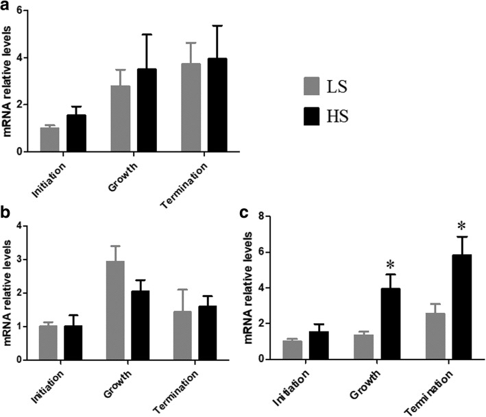
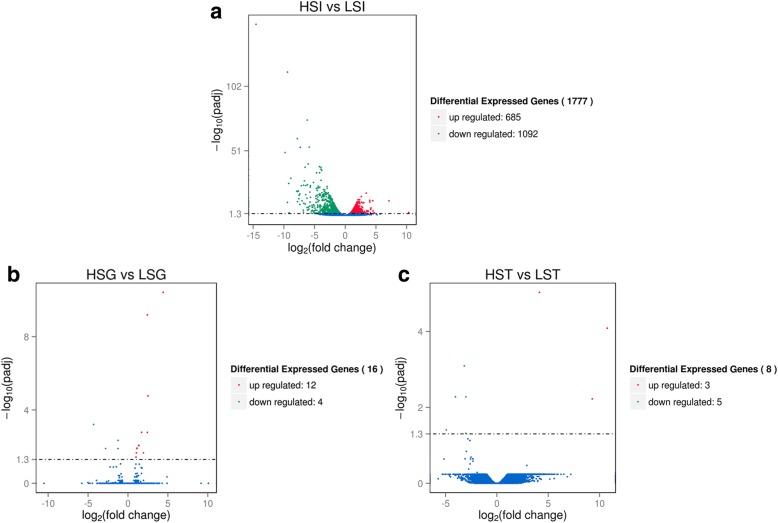
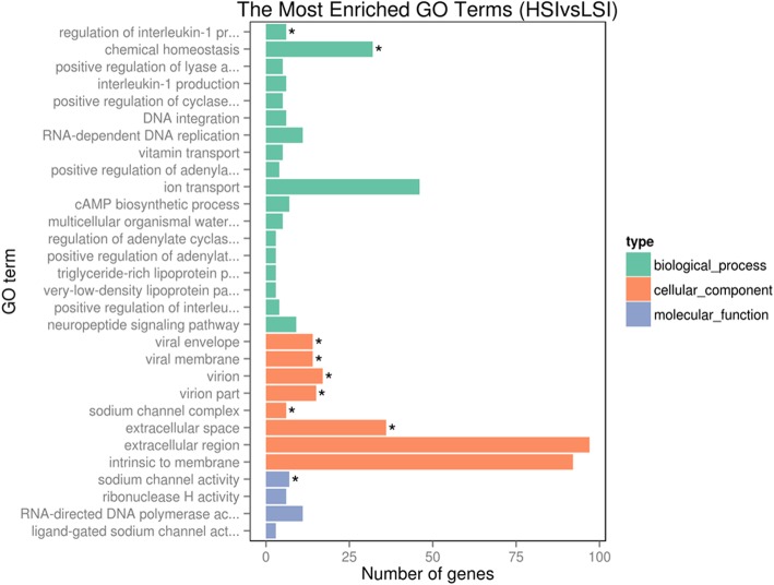
Results: 60 Hy-line Brown laying hens were selected and divided into two groups according to eggshell breaking strength. Eggshell breaking strength of 44.57 ± 0.91 N and 26.68 ± 0.38 N were considered to be the high strength group (HS) and low strength group (LS), respectively. The results showed that mammillary thickness and mammillary knob width of eggshells were significantly lower in the HS. Serum progesterone (P4) and 1,25-dihydroxy vitamin D3 [1,25-(OH)2D3] were significantly higher in the HS compared to the LS during the initiation period of calcification. Serum estradiol (E2) and calcium did not change significantly. All factors mentioned above had no significant differences in the growth and termination periods of calcification. The relative expression of CaBP-D28k and PMCA 1b were not significantly different between HS and LS. The relative expression of NCX1 was significantly higher in HS compared to LS. Moreover, 1777 differentially expressed genes (DEGs) were obtained in the initiation period of calcification. However, few DEGs were identified in the growth or termination periods of calcification. 30 DEGs were selected as candidate genes involved in eggshell calcification during the initiation period of calcification by the analysis of GO terms and KEGG pathways.
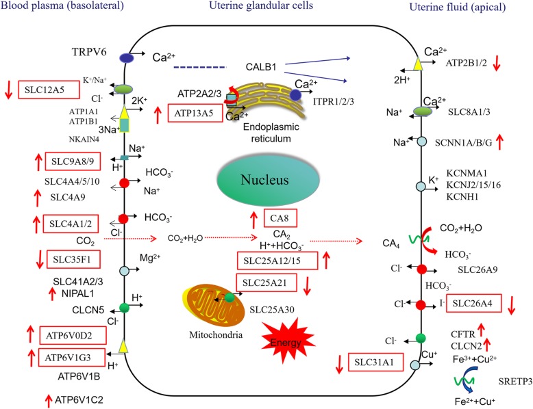
Conclusions: Our study concluded that mammillary thickness and mammillary knob width of the HS were significantly lower than LS. P4 and 1,25-(OH)2D3 were significantly higher in the initiation period of HS. They may impact initial calcification when the mammillary layer is formed. The initiation period of calcification determined eggshell strength rather than the growth or termination periods. We inferred P4 or 1,25-(OH)2D3 may effect the ultrastructure of the mammillary layer by regulating the expression of uterine genes.
Keywords: Eggshell; Gonadal hormone; Ion transport; Laying hen; Transcriptome.
Conflict of interest statement
The authors declare that they have no competing interest.
Figures
Similar articles
-
Uterine transcriptome analysis reveals mRNA expression changes associated with the ultrastructure differences of eggshell in young and aged laying hens.BMC Genomics. 2020 Nov 9;21(1):770. doi: 10.1186/s12864-020-07177-7. BMC Genomics. 2020. PMID: 33167850 Free PMC article.
-
The characterization of uterine calcium transport and metabolism during eggshell calcification of hens laying high or low breaking strength eggshell.Poult Sci. 2025 Jun;104(6):105111. doi: 10.1016/j.psj.2025.105111. Epub 2025 Mar 31. Poult Sci. 2025. PMID: 40222347 Free PMC article.
-
Dietary manganese supplementation modulated mechanical and ultrastructural changes during eggshell formation in laying hens.Poult Sci. 2017 Aug 1;96(8):2699-2707. doi: 10.3382/ps/pex042. Poult Sci. 2017. PMID: 28482094
-
Eggshell color in brown-egg laying hens - a review.Poult Sci. 2015 Oct;94(10):2566-75. doi: 10.3382/ps/pev202. Epub 2015 Aug 3. Poult Sci. 2015. PMID: 26240390 Free PMC article. Review.
-
Effect of genotype and some shell quality traits on lysozyme content and activity in the albumen of eggs from hens under the biodiversity conservation program.Poult Sci. 2021 Mar;100(3):100863. doi: 10.1016/j.psj.2020.11.040. Epub 2020 Nov 27. Poult Sci. 2021. PMID: 33516470 Free PMC article. Review.
Cited by
-
Multiomic analysis revealed the regulatory role of the KRT14 gene in eggshell quality.Front Genet. 2022 Sep 22;13:927670. doi: 10.3389/fgene.2022.927670. eCollection 2022. Front Genet. 2022. PMID: 36212119 Free PMC article.
-
Uterine transcriptome analysis reveals mRNA expression changes associated with the ultrastructure differences of eggshell in young and aged laying hens.BMC Genomics. 2020 Nov 9;21(1):770. doi: 10.1186/s12864-020-07177-7. BMC Genomics. 2020. PMID: 33167850 Free PMC article.
-
The inositol 1,4,5-trisphosphate receptor type 2 protein domains regulate calcium levels and Ion transport gene expression in laying ducks' uterus.Poult Sci. 2025 Jul 3;104(10):105520. doi: 10.1016/j.psj.2025.105520. Online ahead of print. Poult Sci. 2025. PMID: 40633316 Free PMC article.
-
Decreased eggshell strength caused by impairment of uterine calcium transport coincide with higher bone minerals and quality in aged laying hens.J Anim Sci Biotechnol. 2024 Mar 4;15(1):37. doi: 10.1186/s40104-023-00986-2. J Anim Sci Biotechnol. 2024. PMID: 38439110 Free PMC article.
-
The characterization of uterine calcium transport and metabolism during eggshell calcification of hens laying high or low breaking strength eggshell.Poult Sci. 2025 Jun;104(6):105111. doi: 10.1016/j.psj.2025.105111. Epub 2025 Mar 31. Poult Sci. 2025. PMID: 40222347 Free PMC article.
References
-
- Roland DA. Egg shell problems: estimates of incidence and economic-impact. Poult Sci. 1988;67(12):1801–1803. doi: 10.3382/ps.0671801. - DOI
Publication types
MeSH terms
Substances
Grants and funding
LinkOut - more resources
Full Text Sources
Research Materials
