

[Primary intraosseous hematopoietic pseudotumor: clinicopathological analysis and 9-year follow-up of 3 cases]
- PMID: 31511212
- PMCID: PMC6765597
- DOI: 10.12122/j.issn.1673-4254.2019.08.08
[Primary intraosseous hematopoietic pseudotumor: clinicopathological analysis and 9-year follow-up of 3 cases]
Abstract
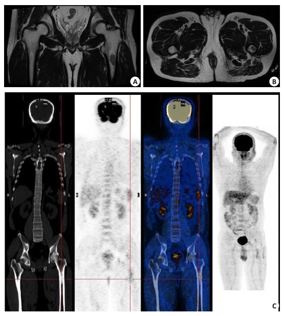
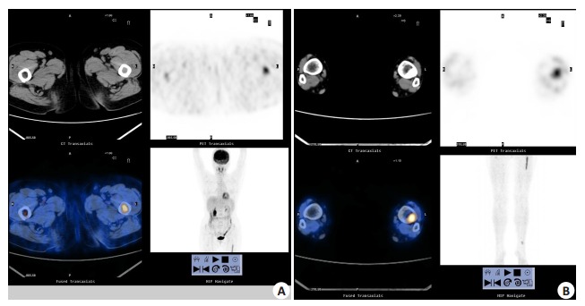
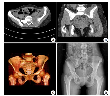
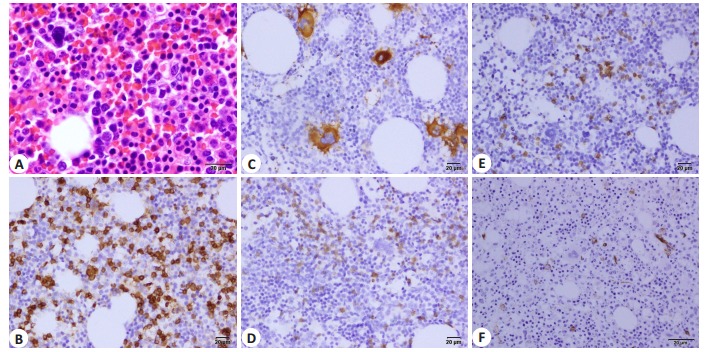
We analyzed the clinicopathological data of 3 cases of primary intraosseous hematopoietic pseudotumor (IHPT), which had been previously misdiagnosed as malignancies or metastases both clinically and pathologically. Two of the patients received close follow-up for 132 and 100 months, and one patient was lost to follow-up, and the tumors were confirmed to be benign in all the 3 cases. IHPT is a rare benign intraosseous solid lesion consisting of tissues resembling normal hematopoietic tissue, and can be easily misdiagnosed as malignancy. Understanding the clinicopathological features and the outcomes of the disease can facilitate the clinical decisions on individualized diagnosis and therapeutic regimens.
本研究收集、分析3例骨内造血细胞假瘤的临床病理资料,初始临床和病理学诊断,都被误诊为恶性肿瘤/转移灶,其中2例分别随访了132月和100月,均证明为良性肿瘤(1例失访)。造血细胞假瘤由类似正常造血组织组成的骨内罕见的良性病变,易被误诊为恶性造血细胞肿瘤,熟知其临床病理特征与转归过程,能够建立正确的个体化诊治方案。
Keywords: extramedullary hematopoiesis; hematopoietic cells hyperplasia; intraosseous hematopoietic pseudotumor.
Figures
Similar articles
-
Focal hematopoietic hyperplasia of the rib--a form of pseudotumor.Skeletal Radiol. 1984;11(2):108-18. doi: 10.1007/BF00348798. Skeletal Radiol. 1984. PMID: 6701544
-
Targeting the Spleen as an Alternative Site for Hematopoiesis.Bioessays. 2019 May;41(5):e1800234. doi: 10.1002/bies.201800234. Epub 2019 Apr 10. Bioessays. 2019. PMID: 30970171 Review.
-
[Comparison of the efficiency of CHOP-based regimen with or without high dose consolidation treatment combined with hematopoietic stem cell transplantation in 63 lymphoblastic lymphoma patients].Zhonghua Zhong Liu Za Zhi. 2009 Jun;31(6):469-73. Zhonghua Zhong Liu Za Zhi. 2009. PMID: 19950562 Chinese.
-
Special Education: Aplastic Anemia.Oncologist. 1996;1(3):187-189. Oncologist. 1996. PMID: 10387986
-
Relationship between Aging and Hematopoietic Cell Transplantation.Biol Blood Marrow Transplant. 2018 Oct;24(10):1965-1970. doi: 10.1016/j.bbmt.2018.08.015. Epub 2018 Aug 18. Biol Blood Marrow Transplant. 2018. PMID: 30130587 Review.
References
-
- Ricchi P, Meloni A, Grigoratos C, et al. Prevalence of extramedullary hematopoiesis, renal cysts, splenic and hepatic lesions, and vertebral hemangiomas among thalassemic patients: a retrospective study from the myocardial iron overload in Thalassemia (MIOT) network. Ann Hematol. 2019;98(6):1333–9. doi: 10.1007/s00277-019-03659-1. - DOI - PubMed
-
- 梁 冬妮, 徐 钢, 李 娟, et al. 以脾脏肿块为首发表现的原发性骨髓纤维化伴髓外造血1例. http://d.old.wanfangdata.com.cn/Periodical/lcysyblxzz201509043 临床与实验病理学杂志. 2015;(9):1075–6.
Publication types
MeSH terms
LinkOut - more resources
Full Text Sources