. 2019 Oct 2;146(20):dev175133.
doi: 10.1242/dev.175133.

Casein kinase 1α decreases β-catenin levels at adherens junctions to facilitate wound closure in Drosophila larvae

Affiliations

Casein kinase 1α decreases β-catenin levels at adherens junctions to facilitate wound closure in Drosophila larvae

Chang-Ru Tsai et al. Development. .

Abstract

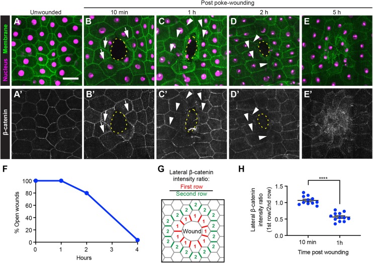
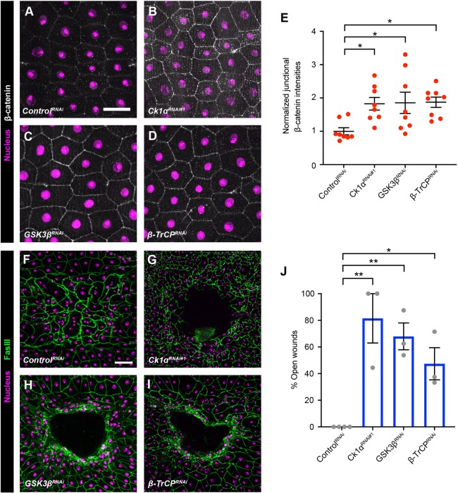
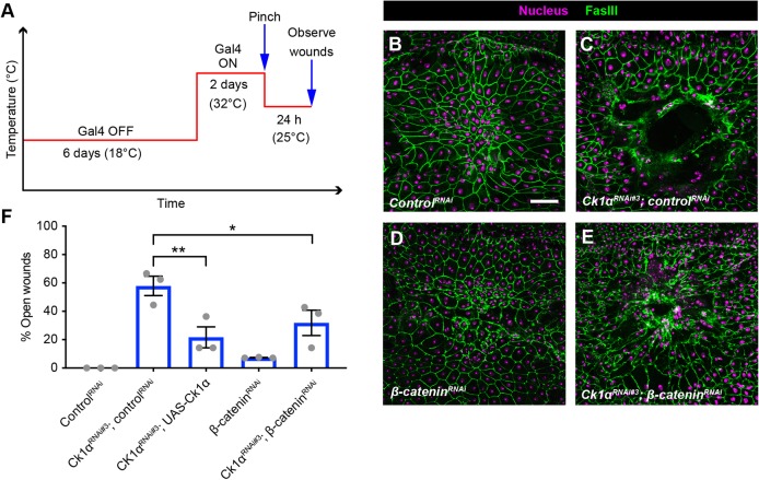
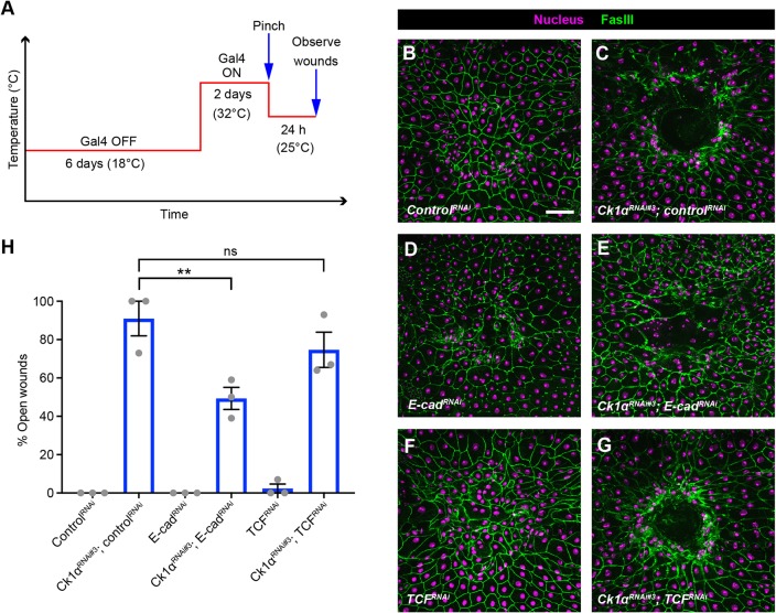
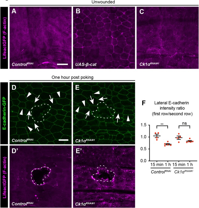
Skin wound repair is essential to restore barrier function and prevent infection after tissue damage. Wound-edge epidermal cells migrate as a sheet to close the wound. However, it is still unclear how cell-cell junctions are regulated during wound closure (WC). To study this, we examined adherens junctions during WC in Drosophila larvae. β-Catenin is reduced at the lateral cell-cell junctions of wound-edge epidermal cells in the early healing stages. Destruction complex components, including Ck1α, GSK3β and β-TrCP, suppress β-catenin levels in the larval epidermis. Tissue-specific RNAi targeting these genes also caused severe WC defects. The Ck1αRNAi -induced WC defect is related to adherens junctions because loss of either β-catenin or E-cadherin significantly rescued this WC defect. In contrast, TCFRNAi does not rescue the Ck1αRNAi -induced WC defect, suggesting that Wnt signaling is not related to this defect. Direct overexpression of β-catenin recapitulates most of the features of Ck1α reduction during wounding. Finally, loss of Ck1α also blocked junctional E-cadherin reduction around the wound. Our results suggest that Ck1α and the destruction complex locally regulate cell adhesion to facilitate efficient wound repair.

Keywords: Adherens junctions; Casein kinase 1α; Drosophila; Epithelium; Wound repair; β-Catenin.


Conflict of interest statement

Competing interestsThe authors declare no competing or financial interests.

Figures

Fig. 1.
Fig. 1.
Junctional β-catenin in wound-edge epidermal cells is reduced after wounding. (A-E′) Dissected epidermal whole mounts of unwounded (A,A′) or poke-wounded (B-E′) third instar larvae expressing UAS-DsRed2nuc (nuclei, magenta) and UAS-src-GFP (cell membranes, green) via the A58-Gal4 driver 10 min (B,B′), 1 h (C,C′), 2 h (D,D′) and 5 h (E,E′) after wounding. (A-E) The nuclei and cell membrane. (A′-E′) The adherens junctions of the same samples immunostained using anti-β-catenin antibodies (white). Scale bar: 50 μm. Dotted yellow lines indicate wound borders. Arrows in B,B′ highlight examples of clear junctional β-catenin signal (B′) and membrane-GFP signal (B). Arrowheads in C-D′ highlight examples of reduced junctional β-catenin (C′,D′) where membrane-GFP is still present (C,D). (F) Quantitation of open poke wounds in control larvae. The epidermal reporter used was e22c-Gal4, UAS-LifeAct-Cherry, UAS-luciferaseRNAi. n≥20 for each time point. (G) Schematic of quantitation strategy for measuring β-catenin levels on lateral segments near the wound. (H) Quantitation of junctional β-catenin ratio in first- and second-row wound-edge epidermal cells. Each dot represents one larva. Data are mean±s.e.m. ****P<0.0001 (unpaired t-test).
Fig. 2.
Fig. 2.
The destruction complex regulates junctional β-catenin levels and wound closure. (A-D) Dissected epidermal whole mounts of unwounded third instar larvae expressing UAS-DsRed2nuc (nuclei, magenta), UAS-src-GFP (cell membranes, green, not shown) and the indicated transgenes via either the e22c-Gal4 (A,C,D) or A58-Gal4 drivers (B). Anti-β-catenin antibody staining is in white. (A) ControlRNAi. (B) Ck1αRNAi#1 (A58-Gal4 used because this line grows slowly with e22c-Gal4). (C) GSK3βRNAi. (D) β-TrCPRNAi. (E) Quantitation of junctional β-catenin intensity in larvae expressing the different transgenes. Each dot represents an average of the β-catenin signal from five junctions of one larva. Data are mean±s.e.m. *P<0.05 (one-way ANOVA). (F-I) Dissected epidermal whole mounts of pinch-wounded third instar larvae expressing UAS-DsRed2Nuc (nuclei, magenta) via the e22c-Gal4 driver and the indicated RNAi transgenes. Cell boundaries were immunostained using anti-Fasciclin III antibodies (green). Scale bars: 50 μm in A for A-D; 100 μm in F for F-I. (F) ControlRNAi. (G) Ck1αRNAi. (H) GSK3βRNAi. (I) β-TrCPRNAi. (J) Quantitation of the percentage of open wounds in larvae expressing the indicated transgenes via the e22c-Gal4 driver. Each dot represents one set of n≥8 larvae for each genotype. Data are mean±s.e.m. *P<0.05, **P<0.01 (one-way ANOVA).
Fig. 3.
Fig. 3.
Silencing β-catenin partially rescues the Ck1αRNAi-induced wound closure defect. (A) Schematic of the experimental design/temperature shift regimen for using Gal80ts to inducibly express UAS-dependent transgenes in the larval epidermis. (B-E) Dissected epidermal whole mounts of pinch-wounded third instar larvae expressing Gal80ts transgene driven by a tubulin promoter, UAS-DsRed2Nuc (nuclei, magenta) via the e22c-Gal4 driver, and the indicated transgenes. Cell boundaries were immunostained using anti-Fasciclin III antibodies (green). (B) ControlRNAi, (C) UAS-Ck1αRNAi#3 and controlRNAi, (D) UAS-β-cateninRNAi, (E) Ck1αRNAi#3 and UAS-β-cateninRNAi. Scale bar: 100 μm. (F) Quantitation of the percentage of open wounds in larvae expressing indicated transgenes via the e22c-Gal4 driver. Each dot represents one set of n≥8 for each genotype. Data are mean±s.e.m. *P<0.05, **P<0.01 (one-way ANOVA).
Fig. 4.
Fig. 4.
The epidermal Ck1αRNAi-induced wound-closure defect is E-cadherin dependent and Wnt signaling independent. (A) Schematic of the experimental design/temperature shift regimen for using Gal80ts to inducibly express UAS-dependent transgenes in the larval epidermis. (B-G) Dissected epidermal whole mounts of pinch-wounded larvae expressing the Gal80ts transgene driven by a tubulin promoter, UAS-DsRed2Nuc (nuclei, magenta) via the e22c-Gal4 driver, and the indicated transgenes 24 h after wounding. Cell boundaries were immunostained using anti-Fasciclin III antibodies (green). (B) ControlRNAi, (C) Ck1αRNAi#3 and controlRNAi, (D) E-cadRNAi, (E) Ck1αRNAi#3 and E-cadRNAi, (F) UAS-TCFRNAi, (G) Ck1αRNAi and UAS-TCFRNAi. Scale bar: 100 μm. (H) Quantitation of the percentage of open wounds in third instar larvae expressing the indicated transgenes via the e22c-Gal4 driver. Each dot represents one set of n≥8 larvae for each genotype. Data are mean±s.e.m. **P<0.01; ns, not significant (one-way ANOVA).
Fig. 5.
Fig. 5.
Epidermal β-catenin overexpression-induced wound closure defect is E-cadherin dependent and Wnt signaling independent. (A) Schematic of the experimental design/temperature shift regimen for using Gal80ts to inducibly express UAS-dependent transgenes in the larval epidermis. (B-G) Dissected epidermal whole mounts of pinch-wounded larvae expressing the Gal80ts transgene driven by a tubulin promoter, UAS-DsRed2Nuc (nuclei, magenta) via the e22c-Gal4 driver, and the indicated transgenes 14 h after wounding. Cell boundaries were immunostained using anti-Fasciclin III antibodies (green). (B) ControlRNAi, (C) UAS-β-catenin and controlRNAi, (D) E-cadRNAi, (E) UAS-β-cat and E-cadRNAi (F), UAS-TCFRNAi, (G) UAS-β-cat and UAS-TCFRNAi. Scale bar: 100 μm. (H) Quantitation of the percentage of open wounds in third instar larvae expressing the indicated transgenes via the e22c-Gal4 driver. Each dot represents one set of n≥8 larvae for each genotype. Data are mean±s.e.m. ***P<0.001; ns, not significant (one-way ANOVA).
Fig. 6.
Fig. 6.
Actin localization and Ck1α are required for the reduction of lateral E-cadherin on wound-edge epidermal cell membranes. (A-C) Dissected larval epidermal whole mounts of third instar larvae expressing UAS-Lifeact-mCherry (magenta) and the indicated transgenes via the e22c-Gal4 driver. (D-E′) Live image of larval epidermis expressing E-cadherin-GFP and UAS-LifeAct-mCherry (magenta) via e22c-Gal4 driver (control, D,D′) and (Ck1αRNAi, E,E′) 1 h after wounding. (D,E) E-cadherin-GFP, green. (D′-E′) F-actin to visualize the wound margins. Arrows and arrowheads in D,E indicate examples of first-row and second-row junctions, respectively. Scale bars: 50 μm. Dotted white lines indicate wound borders. (F) Quantitation of junctional E-cadherin-GFP ratio in first- and second-row wound-edge epidermal cells. Each dot represents one animal. Data are mean±s.e.m. **P<0.01; ns, not significant (unpaired t-test).

Similar articles

Cited by

References

    1. Aberle H., Bauer A., Stappert J., Kispert A. and Kemler R. (1997). beta-catenin is a target for the ubiquitin-proteasome pathway. EMBO J. 16, 3797-3804. 10.1093/emboj/16.13.3797 - DOI - PMC - PubMed
    1. Abreu-Blanco M. T., Verboon J. M., Liu R., Watts J. J. and Parkhurst S. M. (2013). Drosophila embryos close epithelial wounds using a combination of cellular protrusions and an actomyosin purse string. J. Cell Sci. 125, 5984-5997. 10.1242/jcs.109066 - DOI - PMC - PubMed
    1. Baek S. H., Cho H. W., Kwon Y.-C., Lee J. H., Kim M. J., Lee H. and Choe K.-M. (2012). Requirement for Pak3 in Rac1-induced organization of actin and myosin during Drosophila larval wound healing. FEBS Lett. 586, 772-777. 10.1016/j.febslet.2012.01.061 - DOI - PubMed
    1. Brand A. H. and Perrimon N. (1993). Targeted gene expression as a means of altering cell fates and generating dominant phenotypes. Development 118, 401-415. - PubMed
    1. Brock A. R., Wang Y., Berger S., Renkawitz-Pohl R., Han V. C., Wu Y. and Galko M. J. (2012). Transcriptional regulation of Profilin during wound closure in Drosophila larvae. J. Cell Sci. 125, 5667-5676. 10.1242/jcs.107490 - DOI - PMC - PubMed

Publication types

MeSH terms

LinkOut - more resources