

Assessment of uteroplacental vascularisation in early first-trimester pregnancy with contrast-enhanced ultrasound and 3D power Doppler angiography: protocol for a prospective, cross-sectional, multicentre and non-randomised open study ("HOPE Study")
- PMID: 31511289
- PMCID: PMC6747665
- DOI: 10.1136/bmjopen-2019-030353
Assessment of uteroplacental vascularisation in early first-trimester pregnancy with contrast-enhanced ultrasound and 3D power Doppler angiography: protocol for a prospective, cross-sectional, multicentre and non-randomised open study ("HOPE Study")
Abstract
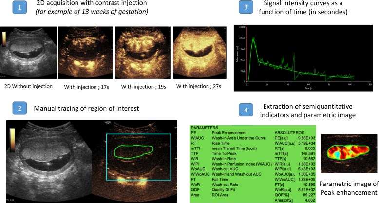
Introduction: Knowledge about the mechanisms leading to the establishment of uteroplacental vascularisation is inadequate, and some of what has been thought to be known for decades has recently been challenged by showing that the intervillous space, the major area of maternal-fetal exchange, appears to be perfused by maternal blood at as early as 6 weeks of gestation. The vascular flow then seems relatively constant until 13 weeks when it appears to increase suddenly.
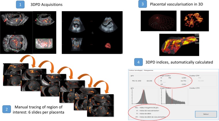
Objectives: The principal objective is to quantify the perfusion of the intervillous space by contrast-enhanced ultrasonography (CEUS) during the first-trimester at three different gestational ages (8, 11 and 13 weeks). The secondary objectives are to: (1) describe the indicators of vascularisation of the placenta (intervillous space) and the myometrium at the three gestational ages, measured by CEUS and three-dimensional power Doppler (3DPD) angiography; (2) compare the diagnostic performance of CEUS and 3DPD for the demonstration and quantification of uteroplacental vascularisation and (3) establish a biological collection of placentas to increase knowledge about placental development and functions during pregnancy.
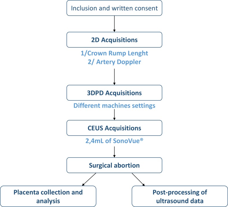
Methods and analysis: This is a prospective, cross-sectional, multicentre and non-randomised open study. We will include 42 women with ongoing pregnancy and divided into three groups of gestational ages (ie, 14 women by per group): 8, 11 and 13 weeks of gestation. 3DPD and then CEUS will be performed and the data about the perfusion kinetics and the 3DPD indices will be calculated and then compared with each other and for each gestational age.
Ethics and dissemination: The appropriate French Ethics Committee Est III approved this study and the related consent forms on 5 April 2016, and the competent authority (Agence Nationale de Sécurité du Médicament et des Produits de Santé) authorised the study on 21 June 2016. The results of this study will be published in a peer-reviewed journal and will be presented at relevant conferences.
Trial registration numbers: ClinicalTrials.gov registry (NCT02884297); EudraCT registry (2015-005655-27).
Keywords: pathology; physiology; prenatal diagnosis; ultrasonography; ultrasound.
© Author(s) (or their employer(s)) 2019. Re-use permitted under CC BY-NC. No commercial re-use. See rights and permissions. Published by BMJ.
Conflict of interest statement
Competing interests: None declared.
Figures
References
Publication types
MeSH terms
Associated data
LinkOut - more resources
Full Text Sources
Medical
Research Materials
Miscellaneous