. 2019 Nov 13;93(23):e01039-19.
doi: 10.1128/JVI.01039-19. Print 2019 Dec 1.

Influenza Viruses in Mice: Deep Sequencing Analysis of Serial Passage and Effects of Sialic Acid Structural Variation

Affiliations

Influenza Viruses in Mice: Deep Sequencing Analysis of Serial Passage and Effects of Sialic Acid Structural Variation

Brian R Wasik et al. J Virol. .

Abstract

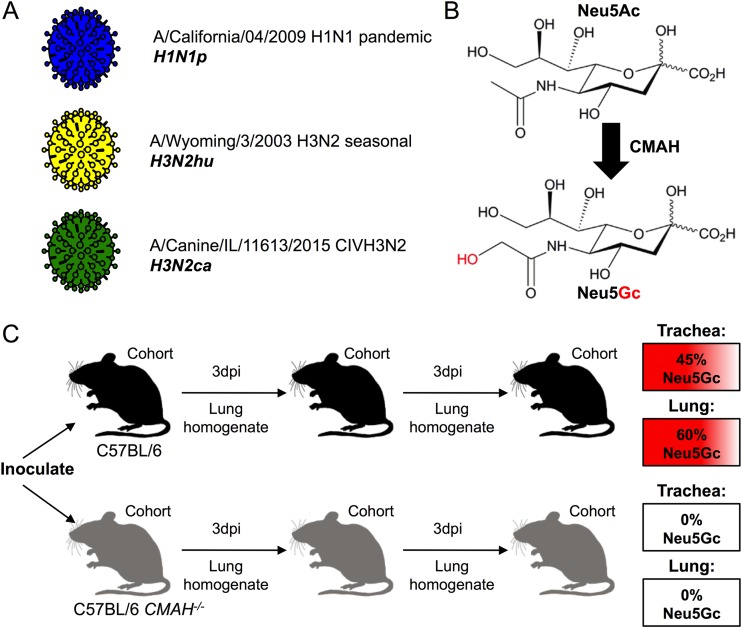
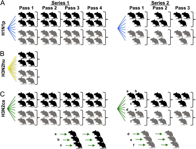
Influenza A viruses have regularly jumped to new host species to cause epidemics or pandemics, an evolutionary process that involves variation in the viral traits necessary to overcome host barriers and facilitate transmission. Mice are not a natural host for influenza virus but are frequently used as models in studies of pathogenesis, often after multiple passages to achieve higher viral titers that result in clinical disease such as weight loss or death. Here, we examine the processes of influenza A virus infection and evolution in mice by comparing single nucleotide variations of a human H1N1 pandemic virus, a seasonal H3N2 virus, and an H3N2 canine influenza virus during experimental passage. We also compared replication and sequence variation in wild-type mice expressing N-glycolylneuraminic acid (Neu5Gc) with those seen in mice expressing only N-acetylneuraminic acid (Neu5Ac). Viruses derived from plasmids were propagated in MDCK cells and then passaged in mice up to four times. Full-genome deep sequencing of the plasmids, cultured viruses, and viruses from mice at various passages revealed only small numbers of mutational changes. The H3N2 canine influenza virus showed increases in frequency of sporadic mutations in the PB2, PA, and NA segments. The H1N1 pandemic virus grew well in mice, and while it exhibited the maintenance of some minority mutations, there was no clear evidence for adaptive evolution. The H3N2 seasonal virus did not establish in the mice. Finally, there were no clear sequence differences associated with the presence or absence of Neu5Gc.IMPORTANCE Mice are commonly used as a model to study the growth and virulence of influenza A viruses in mammals but are not a natural host and have distinct sialic acid receptor profiles compared to humans. Using experimental infections with different subtypes of influenza A virus derived from different hosts, we found that evolution of influenza A virus in mice did not necessarily proceed through the linear accumulation of host-adaptive mutations, that there was variation in the patterns of mutations detected in each repetition, and that the mutation dynamics depended on the virus examined. In addition, variation in the viral receptor, sialic acid, did not affect influenza virus evolution in this model. Overall, our results show that while mice provide a useful animal model for influenza virus pathology, host passage evolution will vary depending on the specific virus tested.

Keywords: animal models; evolution; host range; influenza; sialic acid.


Figures

FIG 1
FIG 1
Outline of the experimental design. (A) Influenza virus stocks were generated from reverse genetic clones: H1N1p (blue), H3N2hu (yellow), and H3N2ca (green). (B) The enzyme CMAH catalyzes the enzymatic conversion of Neu5Ac to Neu5Gc. Inactivation of the cmah gene in a C57BL/6 mouse background generates a Neu5Ac-only mouse. (C) Experimental passages of viruses were performed by nasal inoculation of culture-derived virus or lung homogenates into control C57BL/6 (black) or CMAH−/− (gray) mouse cohorts, 3-day incubation, and harvest of lung homogenates. C57BL/6 mice display Neu5Gc, as a proportion of total Sia, at 45% in trachea and 60% in the lungs. CMAH−/− mice contain no Neu5Gc in their trachea or lungs.
FIG 2
FIG 2
Diagram of experimental mouse passages performed in this study with H1N1p (A), H3N2hu (B), and H3N2ca (C) or with H3N2ca in mouse-to-mouse lineages (C). Individual mice are displayed as control C57BL/6 (black) or CMAH−/− (gray) mice within cohorts. The experiment was performed in two different iterations: series 1 proceeded for four passages among cohorts of four mice (two male, two female), while series 2 proceeded for three passages among cohorts of three mice (an alternating 2:1 sex ratio). H3N2ca mouse lung homogenates from passage 1 of series 2 were also used to initiate a series of mouse-to-mouse lineages in C57BL/6 (a, b, and c) or CMAH−/− (d, e, and f) individual mice.
FIG 3
FIG 3
General influenza virus dynamics in mouse cohorts inoculated with H1N1p (A), H3N2hu (B), or H3N2ca (C). Control C57BL/6 mouse samples are denoted as squares while CMAH−/− mouse samples are shown as triangles. Top, quantitation of influenza virus genome copies (per RT-qPCR of M segment) for each virus was measured for stock virus and pooled lung homogenate (normalized to inoculum volume) to measure genomic bottleneck size at each passage. H3N2hu-inoculated mice lacked measurable genome copies in their lungs at first passage. Bottom, mouse weights were recorded during the course of infection. Figures are from the first passage of series 1 as a representative example. All virus-specific weight-loss phenotypes persisted during the course of each experimental passage and between repeat passage series. Only mice inoculated with H1N1p showed weight loss during the course of infection, with no significant variation between experimental groups.
FIG 4
FIG 4
(A) Expression of the α2,3- and α2,6-linked Sias in the trachea and lungs of wild-type C57BL/6 mice, which express ∼45 to 60% Neu5c, or CMAH−/− mice, which lack Neu5Gc. (B) Examples showing the viral infection in the lungs of experimentally passaged mice by immunohistochemistry for IAV antigen (NP), stained in red. Bar, 100 μm.
FIG 5
FIG 5
Whole-genome deep sequencing metrics and quality control. (A) Gel migration of products of IAV whole-genome RT-PCR shows specific bands of 8 genome segments. Products were present only in infected lung homogenates (+) and absent in uninfected controls (−). RT-PCR mixtures generally contained >100,000 input genome copies. (B) Reads across genome segments were not equal, with frequent bias toward smaller segments. (C) Median reads across the genome segments show some variation in coverage across PB2/PB1/PA segments. Blue line, H1N1p; green line, H3N2ca.
FIG 6
FIG 6
Mutational frequency during H1N1p experimental passage in pooled mouse groups. Single nucleotide variants (SNVs) are represented along the genome segments for the plasmid and virus stocks (black stars), C57BL/6 mice of series 1 (blue circles), CMAH−/− mice of series 1 (blue squares), C57BL/6 mice of series 2 (orange triangles), and CMAH−/− mice of series 2 (orange diamonds). SNVs present at >20% are annotated for their resulting protein sequence changes.
FIG 7
FIG 7
Mutational frequencies during H3N2ca experimental passage in pooled mouse groups. Single nucleotide variants (SNVs) are represented along the genome segments for the plasmid and virus stocks (black stars), C57BL/6 mice of series 1 (green circles), CMAH−/− mice of series 1 (green squares), C57BL/6 mice of series 2 (black triangles), and CMAH−/− mice of series 2 (black diamonds). SNVs present at >20% are annotated for their resulting protein sequence changes.
FIG 8
FIG 8
Mutational frequency at the conclusion (passage 3) of H3N2ca mouse-to-mouse experimental passages in C57BL/6 or CMAH−/− mice. Single nucleotide variants (SNVs) are represented along the genome segments for C57BL/6 lineages a (green circles), b (green squares), and c (green hexagons) and CMAH−/− lineages d (black triangles), e (black diamonds), and f (black inverted triangles). SNVs present at >20% are annotated for their resulting protein sequence changes. FS, frameshift.

References

    1. Longdon B, Brockhurst MA, Russell CA, Welch JJ, Jiggins FM. 2014. The evolution and genetics of virus host shifts. PLoS Pathog 10:e1004395. doi:10.1371/journal.ppat.1004395. - DOI - PMC - PubMed
    1. Subbarao K. 2019. The critical interspecies transmission barrier at the animal-human interface. Trop Med Infect Dis 4:E72. doi:10.3390/tropicalmed4020072. - DOI - PMC - PubMed
    1. Wasik BR, de Wit E, Munster V, Lloyd-Smith JO, Martinez-Sobrido L, Parrish CR. 2019. Onward transmission of viruses: how do viruses emerge to cause epidemics after spillover? Philos Trans R Soc Lond B Biol Sci 374:20190017. doi:10.1098/rstb.2019.0017. - DOI - PMC - PubMed
    1. Yoon S-W, Webby RJ, Webster RG. 2014. Evolution and ecology of influenza A viruses. Curr Top Microbiol Immunol 385:359–375. doi:10.1007/82_2014_396. - DOI - PubMed
    1. Mostafa A, Abdelwhab EM, Mettenleiter TC, Pleschka S. 2018. Zoonotic potential of influenza A viruses: a comprehensive overview. Viruses 10:497. doi:10.3390/v10090497. - DOI - PMC - PubMed

Publication types

MeSH terms

Substances