. 2019 Sep 11;11(509):eaaw8412.
doi: 10.1126/scitranslmed.aaw8412.

Off-target toxicity is a common mechanism of action of cancer drugs undergoing clinical trials

Affiliations

Off-target toxicity is a common mechanism of action of cancer drugs undergoing clinical trials

Ann Lin et al. Sci Transl Med. .

Abstract

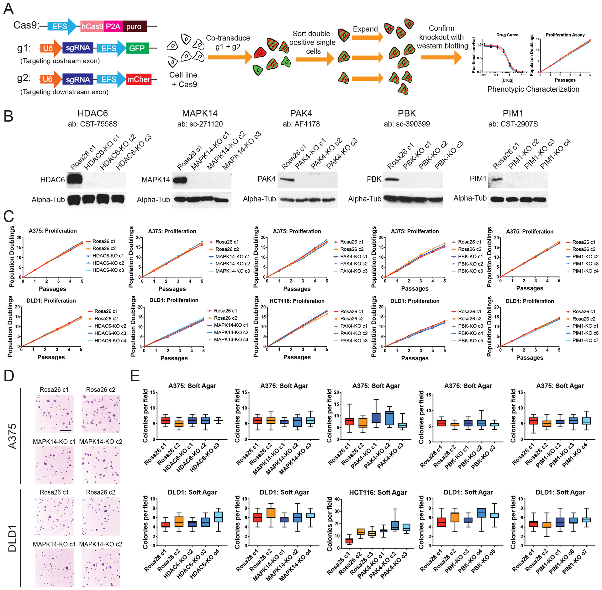
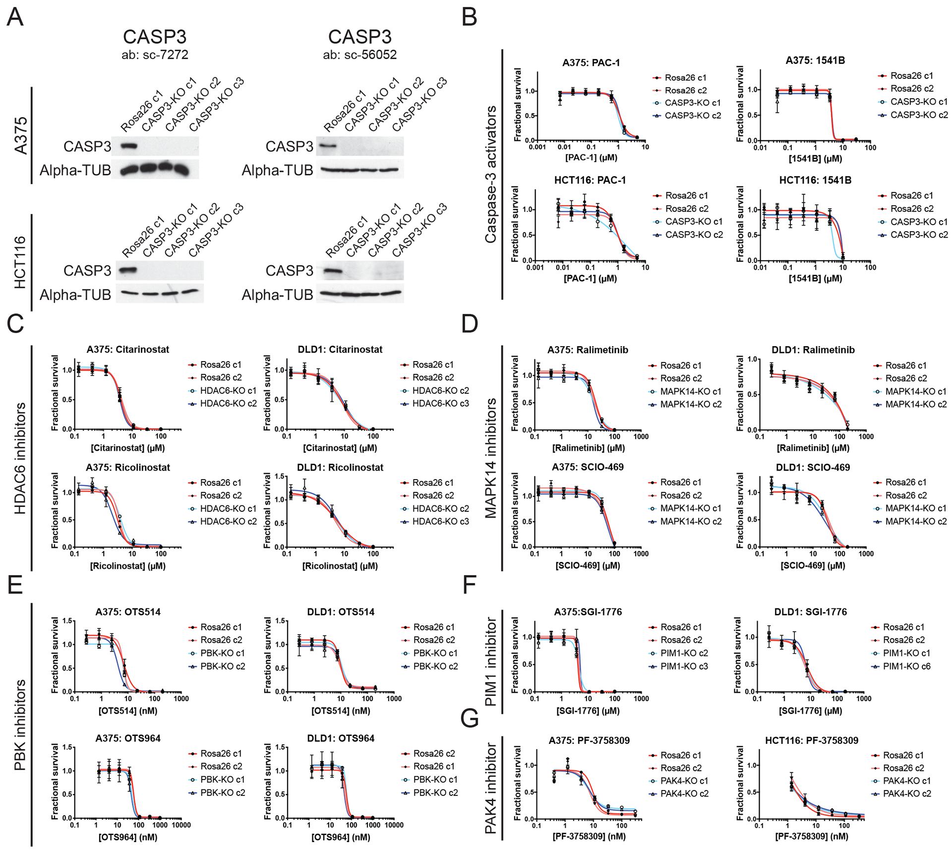
Ninety-seven percent of drug-indication pairs that are tested in clinical trials in oncology never advance to receive U.S. Food and Drug Administration approval. While lack of efficacy and dose-limiting toxicities are the most common causes of trial failure, the reason(s) why so many new drugs encounter these problems is not well understood. Using CRISPR-Cas9 mutagenesis, we investigated a set of cancer drugs and drug targets in various stages of clinical testing. We show that-contrary to previous reports obtained predominantly with RNA interference and small-molecule inhibitors-the proteins ostensibly targeted by these drugs are nonessential for cancer cell proliferation. Moreover, the efficacy of each drug that we tested was unaffected by the loss of its putative target, indicating that these compounds kill cells via off-target effects. By applying a genetic target-deconvolution strategy, we found that the mischaracterized anticancer agent OTS964 is actually a potent inhibitor of the cyclin-dependent kinase CDK11 and that multiple cancer types are addicted to CDK11 expression. We suggest that stringent genetic validation of the mechanism of action of cancer drugs in the preclinical setting may decrease the number of therapies tested in human patients that fail to provide any clinical benefit.


Conflict of interest statement

Competing interests. The authors declare that they have no competing interests.

Figures

Figure 1.
Figure 1.. Cell competition assays to test the essentiality of putative cancer dependencies.
(A) Schematic of the CRISPR-based cell competition assays used in this paper (18). (B) Cell competition assays comparing guides targeting AAVS1 and ROSA26 (non-essential, negative control genes), RPA3 and PCNA (pan-essential positive control proteins), and Aurora A, Aurora B, and ERCC3 (inhibitor-validated cancer dependencies). Full results from these competition experiments are included in data file S2. (C) Cell competition assays for the cell type-specific cancer dependencies BRAF and ESR1. (D) Western blot analysis of A375 populations transduced with the indicated guide RNAs. (E) Cell competition assays with guide RNAs targeting HDAC6, MAPK14, PAK4, PBK, or PIM1 in four different cancer cell lines.
Figure 2.
Figure 2.. Generating and analyzing single cell-derived knockout clones of putative cancer dependencies.
(A) Schematic of the two-guide strategy used to generate clonal knockout cell lines. (B) Western blot analysis of single-cell derived A375 knockout clones. (C) Proliferation assays for HDAC6, MAPK14, PAK4, PBK, and PIM1 knockout clones. (D) Representative images of A375 and DLD1 Rosa26 or MAPK14-KO clones grown in soft agar. Scale bar, 2 mm. (E) Quantification of colony formation in control or knockout A375, DLD1, and HCT116 clones. Boxes represent the 25th, 50th, and 75th percentiles of colonies per field, and the whiskers represent the 10th and 90th percentiles. For each assay, colonies were counted in at least 15 fields under a 10x objective.
Figure 3.
Figure 3.. Target-independent cell killing by multiple anti-cancer drugs.
(A) Western blot analysis for caspase-3 in A375 and HCT116 cells. (B) 7-point dose-response curves of Rosa26 and CASP3-KO A375 and HCT116 cells in the presence of two putative caspase-3 activators, 1541B and PAC-1. (C) 7-point dose-response curves of Rosa26 and HDAC6-KO A375 and DLD1 cells in the presence of two putative HDAC6 inhibitors, ricolinostat and citarinostat. (D) 7-point dose-response curves of Rosa26 and MAPK14-KO A375 and DLD1 cells in the presence of two putative MAPK14 inhibitors, ralimetinib and SCIO-469. (E) 7-point dose-response curves of Rosa26 and PBK-KO A375 and DLD1 cells in the presence of two putative PBK inhibitors, OTS514 and OTS964. (F) 7-point dose-response curves of Rosa26 and PIM1-KO A375 and DLD1 cells in the presence of a putative PIM1 inhibitor, SGI-1776. (G) 7-point dose-response curves of Rosa26 and PAK4-KO A375 and HCT116 cells in the presence of a putative PAK4 inhibitor, PF-3758309.
Figure 4.
Figure 4.. Discovery of CDK11 as the in cellulo target of the mis-characterized anti-cancer drug OTS964.
(A) A schematic of the strategy to use the highly mutagenic HCT116 cell line to isolate mutations that confer OTS964 resistance. (B) Sanger sequencing validation of two heterozygous mutations in the CDK11B kinase domain. (C) Constructs used to introduce the G579S mutation into CDK11B via CRISPR-mediated HDR. The yellow arrowhead indicates the site of Cas9 cleavage, the red bar indicates the G579S substitution, and the blue bars indicate silent mutations introduced to prevent re-cutting after HDR. (D) Crystal violet staining of cancer cells transfected with the indicated constructs and then cultured in a lethal concentration of OTS964. (E) 7-point dose-response curves of Rosa26, PBK-KO, and CDK11BG579S clones grown in varying concentration of OTS964. (F) Titration experiments reveal that OTS964 binds to CDK11B with a KD of 40 nM. (G) Pancreatic cancer cell line MiaPaca-2 was transduced with guides specific CDK11A, guides specific for CDK11B, or guides that harbored cut sites in both genes. (H) A375 H2B-mCherry cells (left) or A375 H2B-mCherry cells that express CDK11BG579S (right) were arrested at G1/S with a double-thymidine block and then were released into normal medium or medium containing OTS964. The percentage of mitotic cells in each population was scored every hour. (I) Representative images of the experiments in (H), 9 hours after release from thymidine. Scale bar, 50 μm.

Comment in

  • Avoiding target misidentification.
    Paiva SL. Paiva SL. Nat Rev Drug Discov. 2019 Oct;18(11):826. doi: 10.1038/d41573-019-00161-1. Nat Rev Drug Discov. 2019. PMID: 31673126 No abstract available.

References

    1. Luo J, Solimini NL, Elledge SJ, Principles of Cancer Therapy: Oncogene and Non-oncogene Addiction, Cell 136, 823–837 (2009). - PMC - PubMed
    1. Sharma SV, Settleman J, Oncogene addiction: setting the stage for molecularly targeted cancer therapy, Genes Dev. 21, 3214–3231 (2007). - PubMed
    1. Lin A, Giuliano CJ, Sayles NM, Sheltzer JM, CRISPR/Cas9 mutagenesis invalidates a putative cancer dependency targeted in on-going clinical trials, eLife 6, e24179 (2017). - PMC - PubMed
    1. Giuliano CJ, Lin A, Smith JC, Palladino AC, Sheltzer JM, MELK expression correlates with tumor mitotic activity but is not required for cancer growth, eLife 7, e32838 (2018). - PMC - PubMed
    1. Wong CH, Siah KW, Lo AW, Estimation of clinical trial success rates and related parameters, Biostatistics (2018), doi:10.1093/biostatistics/kxx069. - DOI - PMC - PubMed

Publication types

MeSH terms