. 2019 Sep 11;10(1):4112.
doi: 10.1038/s41467-019-12013-y.

In vivo epigenetic editing of Sema6a promoter reverses transcallosal dysconnectivity caused by C11orf46/Arl14ep risk gene

Affiliations

In vivo epigenetic editing of Sema6a promoter reverses transcallosal dysconnectivity caused by C11orf46/Arl14ep risk gene

Cyril J Peter et al. Nat Commun. .

Abstract

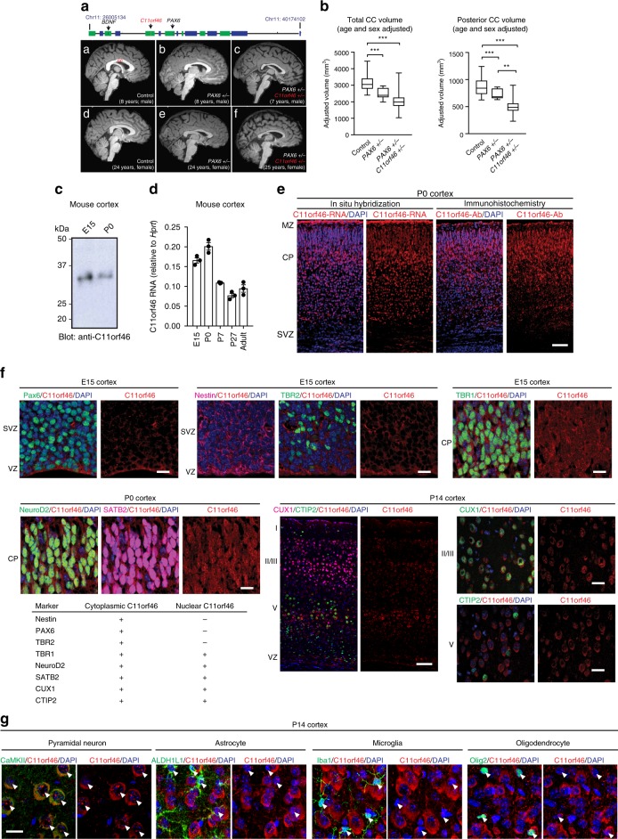
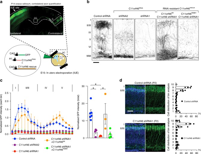
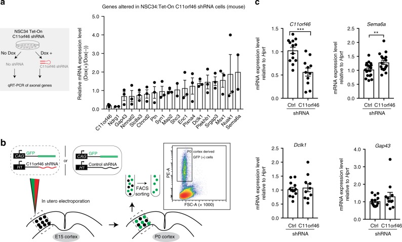
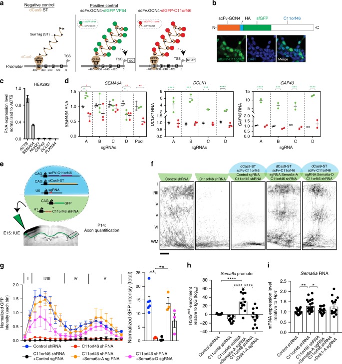
Many neuropsychiatric risk genes contribute to epigenetic regulation but little is known about specific chromatin-associated mechanisms governing the formation of neuronal connectivity. Here we show that transcallosal connectivity is critically dependent on C11orf46, a nuclear protein encoded in the chromosome 11p13 WAGR risk locus. C11orf46 haploinsufficiency was associated with hypoplasia of the corpus callosum. C11orf46 knockdown disrupted transcallosal projections and was rescued by wild type C11orf46 but not the C11orf46R236H mutant associated with intellectual disability. Multiple genes encoding key regulators of axonal development, including Sema6a, were hyperexpressed in C11orf46-knockdown neurons. RNA-guided epigenetic editing of Sema6a gene promoters via a dCas9-SunTag system with C11orf46 binding normalized SEMA6A expression and rescued transcallosal dysconnectivity via repressive chromatin remodeling by the SETDB1 repressor complex. Our study demonstrates that interhemispheric communication is sensitive to locus-specific remodeling of neuronal chromatin, revealing the therapeutic potential for shaping the brain's connectome via gene-targeted designer activators and repressor proteins.


Conflict of interest statement

J.C.H. is the recipient of an unrestricted research grant from Rhythm Pharmaceuticals. The other authors declare no competing interests.

Figures

Fig. 1
Fig. 1
C11orf46 is a neuronal nuclear protein important for callosal development. a (top) Genomic map of chr. 11 WAGR deletion locus. (A–F) Representative sagittal T1 MRI images from midline were shown. A) 8-year-old male control; B) 8-year-old male isolated PAX6+/−; C) 7-year-old male heterozygous 11p13 deletion with PAX6+/− and C11orf46+/−; D) 24-year-old female control; E) 24-year-old female isolated PAX6+/−; F) 25-year-old female heterozygous 11p13 deletion with PAX6+/− and C11orf46+/−. b Hypoplastic corpus callosum (CC) in patients with 11p13 deletion encompassing C11orf46. ANCOVA including age and sex as covariates, compared CC volumes of control (n = 23), isolated PAX6+/− (n = 12), and heterozygous 11p13 deletion with PAX6+/− and C11orf46+/− (n = 27). P-values are shown from two-sided post hoc LSD comparisons: **P < 0.01, ***P < 0.001. Box and whisker plots represent the distribution of unadjusted CC volume. The inner bar indicates median value. The upper and lower box ends represent first and third quantile. The upper and lower whisker ends represent the maximum and minimum values. c C11orf46 protein in mouse cortices at embryonic day 15 (E15) and postnatal day 0 (P0). d mRNA expression of C11orf46 in the mouse cerebral cortex during prenatal and postnatal development (mean ± SEM). n = 3. e (Left) C11orf46 mRNA (red) is predominantly expressed in the cortical plate (CP) in the somatosensory cortex at P0. (Right) C11orf46 protein (red) is predominantly expressed in the CP at P0. Blue, nucleus. Scale bar, 50 µm. MZ, marginal zone; SVZ, subventricular zone. f C11orf46 (red) is expressed in the cytoplasm, but not in the nucleus in Nestin-, PAX6-, and TBR2-positive cells, whereas C11orf46 is expressed in the cytoplasm and in the nucleus with a punctate pattern in TBR1-, NeuroD2-, SATB2-, CUX1-, and CTIP2-positive neuronal cells. (bottom left) Cell type specific C11orf46 expression are summarized in the table. Scale bar, 10 and 100 μm (low magnification images). g Representative images of C11orf46 protein staining with cell type-specific markers (white arrowheads) at P14. C11orf46 is predominantly expressed in CaMKII-positive neurons, but not Olig2 (oligodendrocyte), ALDH1L1 (astrocyte), Iba-1 (microglia) -positive cells. Scale bar, 20 μm
Fig. 2
Fig. 2
Suppression of C11orf46 impairs callosal development and interhemispheric connectivity. a (top) Schematic representation of in utero electroporation of GFP expressing plasmid into somatosensory cortex at E15. (bottom) P14 somatosensory cortex expressing GFP in pyramidal neurons at IUE injection site (Ipsilateral side) and axonal projections between layer I–VI (Contralateral side). b Knockdown of C11orf46 in pyramidal neurons of layer II/III (Ipsilateral) show impaired axonal terminal arborization in the contralateral somatosensory cortex at P14. C11orf46 shRNA1 had a stronger axonal arborization impairment than shRNA2 when compared with control shRNA (first 3 panels); this axonal deficit was partially rescued by co-expressing RNAi resistant wild-type C11orf46 (C11orf46Wt), but not by R236H mutant (C11orf46R236H) (last 2 panels). Scale bar, 100 µm. c Layer distributions (left) and total axonal density (right) of callosal axons in the contralateral site of the electroporated brains were shown as normalized immunofluorescence intensities of callosal axons. F (4, 15) = 7.9591, P = 0.0012 was determined by one-way ANOVA with post hoc Bonferroni test. Three to six mice per condition. *P < 0.05. Impaired axon terminal arborization across all cortical layers elicited by C11orf46 knockdown was restored by overexpression of C11orf46Wt, but not by overexpression of C11orf46R236H. d C11orf46 knockdown did not have an effect on radial neuronal migration at P2. Scale bar, 50 μm. Bar graphs indicate mean ± S.E.M (c, d)
Fig. 3
Fig. 3
C11orf46 binds to the SETDB1 complex. a Flow diagrams for immunoaffinity purification of C11orf46 followed by mass spectrometric analysis. b Eight clones of inducible HEK293 cell lines expressing FLAG-tagged C11orf46 (clone 1–5 and 10) and a truncated form C11orf46 lacking amino acids 209–237 at the C-terminal region which corresponds to cysteine rich domain (CRD) of C11orf46 (C11orf46∆) (clone 7 and 8) in the presence of doxycycline (Dox+). SETDB1 was co-precipitated with C11orf46, but not with C11orf46∆. c Silver staining of proteins co-precipitated with C11orf46 (clone 10) or C11orf46∆ (clone 8). SETDB1, MCAF1 and KAP1 were detected in co-precipitate from C11orf46, but not of C11orf46∆. d Top scoring proteins co-precipitated with C11orf46 in cell lines overexpressed with C11orf46, C11orf46∆, and control cells are shown with % of amino acid sequence coverage. e Protein abundance in affinity purified C11orf46 complexes. Notice prominence of SETDB1-MCAF1-KAP1 repressor proteins (black). Bar graphs indicate mean ± S.E.M. n = 3 independent quantification. f Schematic diagrams indicating position of C11orf46 arginine to histidine substitution at codon 236 (R236H) in conserved cysteine rich domain (CRD; amino acids 209–237) between human and mouse. g, h Co-immunoprecipitation experiments using protein lysates from HEK293 cells overexpressing FLAG-tagged C11orf46, C11orf46∆, or C11orf46R236H showed that absence of CRD domain and R236H mutation is associated with weakened or non-detectable C11orf46-SETDB1 binding and partial loss of binding to other components. R236H mutation does not fully disrupt C11orf46-KAP1 binding. i R236H mutation and CRD deletion of C11orf46 do not affect binding to histone H3. j Flow diagrams for immunoaffinity purification of SETDB1 followed by mass spectrometric analysis. k Inducible SETDB1 expression system in HEK293 cell lines (48 h after Dox treatment). l, m Silver staining of proteins co-precipitated with SETDB1. Both endogenous C11orf46 and SETDB1 binding partners were detected by mass spectrometry. Top scoring proteins co-precipitated with SETDB1 in cell lines overexpressed with SETDB1 and control cells are shown. n (left) C11orf46-SETDB1 co-immunoprecipitation in human cerebral cortex. (right) SETB1 co-immunoprecipitates include C11orf46, HP1γ, and MCAF1 in HeLa nuclear extract. o Schematic summary of above data representing working model of C11orf46-SETDB1 complex in relation to histone H3
Fig. 4
Fig. 4
Selection of targeted genes by C11orf46-SETDB1 complex. a Mouse neuroblastoma cells NSC34 inducibly expressing C11orf46 shRNA upon doxycycline addition (schematic in left panel); selected subset of 17 differentially expressed genes implicated in axonal growth and development was tested in RNA expressed from NSC34:Tet-On C11orf46 shRNA cell (right panel). b Neuronal progenitors received control or C11orf46 shRNA and GFP marker plasmids at somatosensory cortex by IUE at E15; four days later at P0 GFP-positive cells were collected by fluorescence-activated cell sorting (FACS). c C11orf46 transcript levels were reduced by 50% in sorted GFP-positive cortical neurons electroporated with C11orf46 shRNA1 as compared to GFP-positive neurons with control shRNA (P = 0.0003). Note increase of Sema6a transcript upon C11orf46 knockdown (P = 0.0085). *P < 0.05, **P < 0.01, ***P < 0.001 determined by Student’s t-test test. n = 10–19 independent mice per condition. All data were normalized by the Hprt transcript. Bar graphs indicate mean ± S.E.M (a, c)
Fig. 5
Fig. 5
C11orf46- epigenomic editing of neurite-regulating genes rescues transcallosal dysconnectivity in C11orf46-deficient neurons. a Schematic representation showing epigenome editing using nuclease-deficient CRISPR/Cas9 SunTag (dCas9-ST) system. Single guide RNA (sgRNA) targeting neuronal gene promoters is introduced with dCas9-ST (10xGCN4) and scFv-sfGFP-C11orf46 (or −VP64 as a positive control) in HEK293 cells. b Overexpressed fGFP-C11orf46 is colocalized with DAPI signals in HEK293 cells. c Relative expression levels of neurite-regulating genes in HEK293 cells. d dCas9-ST mediated promoter loading with 10xC11orf46, and C11orf46R236H in comparison to 10xVP64 at SEMA6A in HEK293 cells. Four sgRNAs (ad) for each gene were tested individually, and for SEMA6A also as a pool. Two-way ANOVA followed by Dunnett’s multiple comparisons test was performed where C11orf46- or VP64-epigenetic editing effects were tested. e Epigenomic editing of transcallosal neurons by IUE. Five transgenes were delivered simultaneously at E15 by IUE into the developing somatosensory cortex, followed by quantification of axonal arborization at P14. f, g Axonal arborization was disrupted by C11orf46 knockdown, and restored by dCas9-ST mediated recruitment of C11orf46 to Sema6a promoter (Sema6a-), using sgRNAs Sema6a-A, Sema6a-D, but not with non-targeting sgRNA. Scale bar, 100 µm. Layer distributions and total axonal density in the contralateral site of the electroporated brains are shown (g). F (4, 13) = 13.1934, P = 0.0002 was determined by one-way ANOVA with post hoc Bonferroni test. Three to six mice per condition. h H3K9me3 enrichment at Sema6a promoter in GFP + neurons at P0, four days after IUE. Epigenome editing by Sema6a-A sgRNA increases H3K9me3 levels at Sema6a promoter. Dclk1-A sgRNA did not affect H3K9me3 levels at Sema6a promoter. F (3,44) = 8.718, P = 0.0001 was determined by one-way ANOVA with post hoc Bonferroni test. i Sema6a mRNA level in GFP + neurons at P0. Increased Sema6a expression caused by C11orf46 knockdown were suppressed by C11orf46-epigenome editing using Sema6a-A sgRNA, but not by Dclk1-A sgRNA. F (3,72) = 4.746, P = 0.0045. *P < 0.05, **P < 0.01 determined by one-way ANOVA with post hoc Bonferroni test. *P < 0.05, **P < 0.01, ***P < 0.001, and ****P < 0.0001. Bar graphs indicate mean ± S.E.M.

Similar articles

Cited by

References

    1. Peiker I, et al. Perceptual integration deficits in autism spectrum disorders are associated with reduced interhemispheric gamma-band coherence. J. Neurosci. 2015;35:16352–16361. doi: 10.1523/JNEUROSCI.1442-15.2015. - DOI - PMC - PubMed
    1. Ribolsi M, Daskalakis ZJ, Siracusano A, Koch G. Abnormal asymmetry of brain connectivity in schizophrenia. Front. Hum. Neurosci. 2014;8:1010. doi: 10.3389/fnhum.2014.01010. - DOI - PMC - PubMed
    1. Margari L, et al. Clinical manifestations in children and adolescents with corpus callosum abnormalities. J. Neurol. 2016;263:1939–1945. doi: 10.1007/s00415-016-8225-x. - DOI - PubMed
    1. Lindwall C, Fothergill T, Richards LJ. Commissure formation in the mammalian forebrain. Curr. Opin. Neurobiol. 2007;17:3–14. doi: 10.1016/j.conb.2007.01.008. - DOI - PubMed
    1. Mita S, et al. Transcallosal projections require glycoprotein M6-dependent neurite growth and guidance. Cereb. Cortex. 2015;25:4111–4125. doi: 10.1093/cercor/bhu129. - DOI - PubMed

Publication types

MeSH terms