. 2019 Sep 11;9(1):13090.
doi: 10.1038/s41598-019-49482-6.

Prion protein modulates endothelial to mesenchyme-like transition in trabecular meshwork cells: Implications for primary open angle glaucoma

Affiliations

Prion protein modulates endothelial to mesenchyme-like transition in trabecular meshwork cells: Implications for primary open angle glaucoma

Ajay Ashok et al. Sci Rep. .

Abstract

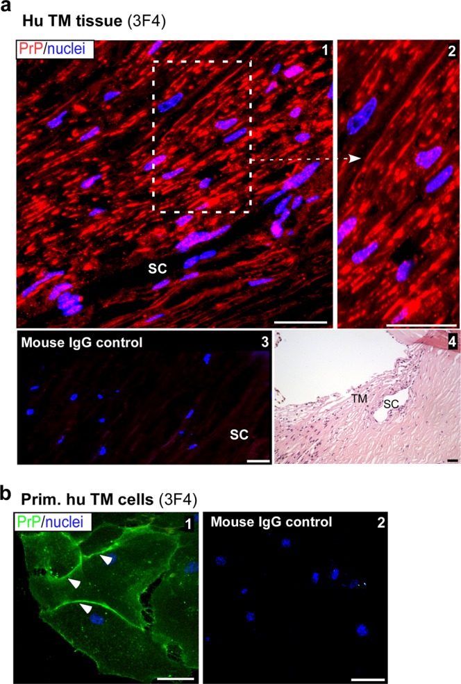
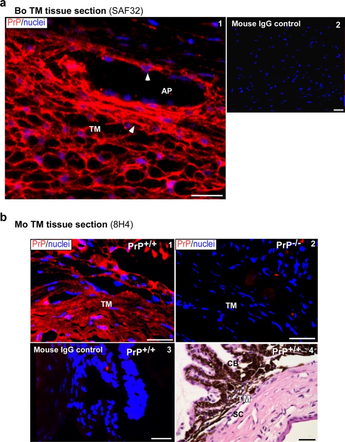
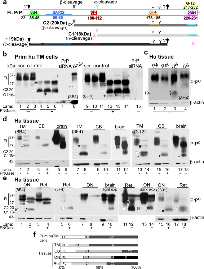
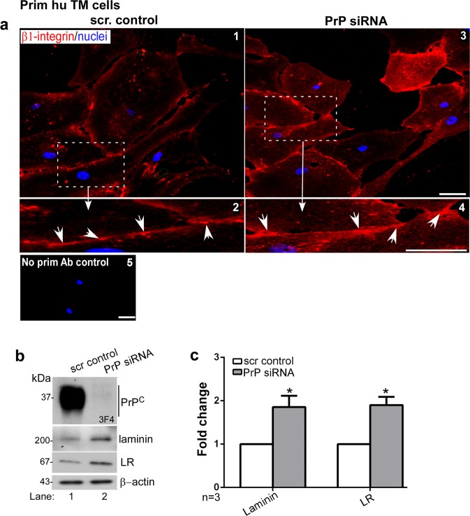
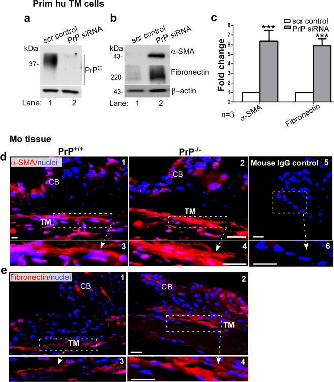
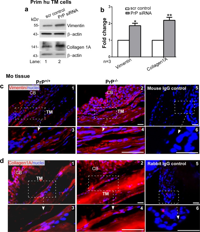
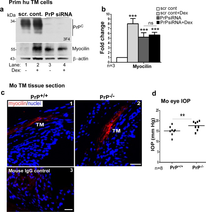
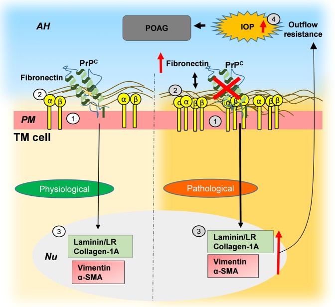
Endothelial-to-mesenchyme-like transition (Endo-MT) of trabecular meshwork (TM) cells is known to be associated with primary open angle glaucoma (POAG). Here, we investigated whether the prion protein (PrPC), a neuronal protein known to modulate epithelial-to-mesenchymal transition in a variety of cell types, is expressed in the TM, and plays a similar role at this site. Using a combination of primary human TM cells and human, bovine, and PrP-knock-out (PrP-/-) mouse models, we demonstrate that PrPC is expressed in the TM of all three species, including endothelial cells lining the Schlemm's canal. Silencing of PrPC in primary human TM cells induces aggregation of β1-integrin and upregulation of α-smooth muscle actin, fibronectin, collagen 1A, vimentin, and laminin, suggestive of transition to a mesenchyme-like phenotype. Remarkably, intraocular pressure is significantly elevated in PrP-/- mice relative to wild-type controls, suggesting reduced pliability of the extracellular matrix and increased resistance to aqueous outflow in the absence of PrPC. Since PrPC is cleaved by members of the disintegrin and matrix-metalloprotease family that are increased in the aqueous humor of POAG arising from a variety of conditions, it is likely that concomitant cleavage of PrPC exaggerates and confounds the pathology by inducing Endo-MT-like changes in the TM.


Conflict of interest statement

The authors declare no competing interests.

Figures

Figure 1
Figure 1
Distribution of PrPC in the human trabecular meshwork. (a) Immunoreaction of human TM section with PrP-specific antibody 3F4 followed by Alexa fluor 546-conjugated secondary antibody shows strong reactivity in all layers of the TM (panel 1). High magnification image demonstrates expression of PrPC on the plasma membrane of TM cells (panel 2). A serial section reacted with mouse IgG and Alexa fluor 546-conjugated secondary antibody shows no reaction (panel 3). H&E staining of a serial section confirms the TM region and Schlemm’s canal (SC) (panel 4). Scale bar: 25 µm. (b) Non-permeabilized primary human TM cells reacted with 3F4 followed by Alexa Fluor 488-conjugated secondary antibody show expression of PrPC on the plasma membrane (panel 1). No reaction is detected in control cells exposed to mouse IgG followed by the same secondary antibody (panel 2). Scale bar: 25 µm.
Figure 2
Figure 2
Expression of PrPC in bovine and murine TM. (a) Immunoreaction of bovine TM section with PrP-specific antibody SAF32 followed by Alexa fluor 546-conjugated secondary antibody shows strong reactivity for PrPC on the plasma membrane of TM cells and endothelial cells lining the aqueous plexus (AP) (panel 1, arrowhead). No reaction is detected in a serial section exposed to mouse IgG followed by the same secondary antibody (panel 2). Scale bar: 25 µm. (b) Immunoreaction of the anterior segment of PrP+/+ mouse eye with PrP-specific antibody 8H4 followed by Alexa fluor 546-conjugated secondary antibody shows expression of PrPC in all layers of the TM (panel 1). No reaction is noted in PrP−/− mouse sample processed in parallel (panel 2). Reaction of PrP+/+ sample with mouse IgG followed by the same secondary antibody shows no reaction (panel 3). H&E staining of a serial section confirms accurate identification of the TM region (panel 4). Scale bar: 25 µm.
Figure 3
Figure 3
Processing of PrPC in human ocular tissue. (a) Schematic representation of full length (FL), α-cleaved (C1, 18 kDa), β-cleaved (C2, 20 kDa), and ~19 kDa forms of PrPC. Antibody 8B4 reacts with FL and N-terminal fragments of PrPC, 3F4 reacts with FL and C2, and 8H4, G-12, and 2301 react with FL, C1, and C2. (b) Probing of lysates from primary human TM cells cultured from three different cases with 3F4 and 8H4 shows FL and mainly C2 fragment of PrPC. C1 represents a small fraction of total PrPC. Human brain lysate provides a positive control, and lysates from cells transfected with PrP-siRNA serve as a negative control (lanes 1–15). (c) Probing of lysates from the TM, retina (Ret), optic nerve (ON), and CB with 8H4 shows glycosylated PrPC in all samples (lanes 1–4), and a ~19 kDa fragment in lysates from the ON (lane 3,?) (d) Probing of human TM and CB lysates with 8B4, 3F4, and G-12 shows FL glycosylated and deglycosylated PrPC in all samples as in human brain. The TM shows significantly more C2 relative to FL and C1, the CB shows mainly C1, while the brain shows mainly FL and a small amount of C1 (lanes 1–18) (lighter exposures are shown for lanes 11 and 12. Complete membrane is shown in Supplementary Fig. S2). TM and CB lysates probed with 2301 antibody mimicked the G-12 probing data (Supplementary Fig. S3) (e) Probing of lysates from the ON and retina with 8B4, 3F4, and 2301 shows FL PrPC as in human brain, and a ~19 kDa fragment in deglycosylated ON sample. The retina has significantly more C2 relative to FL and C1, while the ON has more C1 relative to C2. The ~19 kDa fragment does not react with 2301 (lanes 15-18). (FL: #; C1: star; C2: white arrowhead;?: ~19 kDa). All membranes were re-probed for β-actin to control for loading. (f) The relative abundance of FL, C1, C2, and the ~19 kDa fragment is shown graphically. Figures 3c-e are from tissue harvested from the same eye. Similar results were obtained from two other eye globes.
Figure 4
Figure 4
Downregulation of PrPC aggregates β1-integrin and upregulates laminin and its receptor. (a) Silencing of PrPC in human TM cells followed by immunostaining with antibody specific for activated β1-integrin shows clustering of activated β1-integrin on the plasma membrane in the absence of PrPC (arrowheads) (panels 1–4). Cells transfected with PrP-siRNA and reacted with mouse IgG and respective secondary antibody do not show any reaction (panel 5). Scale bar: 25 µm. (b) Probing of TM cell lysates for PrPC shows the expected glycoforms in control cells transfected with scrambled siRNA, and minimal reaction in cells exposed to PrP-siRNA (lanes 1 & 2). Probing for laminin and laminin receptor (LR) shows significant upregulation in the absence of PrPC relative to control (lanes 1 & 2). (c) Quantification by densitometry after normalization with β-actin shows 2-fold upregulation of laminin and LR due to downregulation of PrPC. Values are mean ± SEM of the indicated n. *p < 0.05. Full-length blots are included in the Supplementary Fig. S2.
Figure 5
Figure 5
Downregulation of PrPC upregulates α-SMA and fibronectin in the TM. (a) Probing of TM cell lysates treated with scrambled and PrP-siRNA for PrPC shows the expected glycoforms in the scrambled control, and minimal reactivity for PrPC in the experimental sample (lanes 1 & 2). (b) Probing of the same lysates for α-SMA and fibronectin shows upregulation in the absence of PrPC relative to controls (lanes 1 & 2). (c) Quantification of protein expression by densitometry after normalization with β-actin shows 6.1-fold and 5.9-fold upregulation of α-SMA and fibronectin respectively. Values are mean ± SEM of the indicated n. *p < 0.05, **p < 0.01. Full-length blots (Supplementary Fig. S2). (d & e) Immunoreaction of fixed sections from the anterior segment of PrP+/+ and PrP−/− mice shows stronger reactivity for α-SMA and fibronectin in the TM of PrP−/− relative to PrP+/+ samples (panel 1 vs. 2). Scale bar: 25 µm. No reaction was detected in samples reacted with mouse IgG followed by Alexa 546-conjugated secondary antibody (panels 5 & 6).
Figure 6
Figure 6
Downregulation of PrPC upregulates vimentin and collagen 1A. (a) PrPC was silenced in human TM cells and lysates were processed as above. Probing for vimentin and collagen 1A shows significant upregulation in the absence of PrPC relative to controls (lanes 1 & 2). (b) Quantification of protein expression by densitometry after normalization with β-actin shows 1.9-fold upregulation of vimentin and 2.2-fold upregulation of collagen 1A in the absence of PrPC. Values are mean ± SEM of the indicated n. *p < 0.05, **p < 0.01. Full-length blots (Supplementary Fig. S2). (c & d) Immunoreaction of fixed sections from the anterior segment of PrP+/+ and PrP−/− mice for vimentin and collagen 1A shows stronger reaction in the TM of PrP−/− relative to PrP+/+ samples (panel 1 vs. 2). No reaction was detected when serial sections were reacted with mouse or rabbit IgG followed by Alexa 546-conjugated secondary antibody (panels 5 & 6). Scale bar: 25 µm.
Figure 7
Figure 7
Myocilin is upregulated by downregulation of PrPC. (a) PrPC was silenced in primary human TM cells and lysates were processed as above. Probing for PrPC shows the expected glycoforms in controls, and minimal reaction in samples treated with PrP-siRNA (lanes 1 & 2 vs. 3 & 4). Re-probing for myocilin shows significant upregulation in the absence of PrPC (lane 1 vs 3). Exposure of control and experimental cells to dexamethasone (Dex) shows upregulation of myocilin as expected (lanes 1 & 2). However, no additive effect of dexamethasone is noted in the absence of PrPC (lanes 3 & 4). (b) Quantification by densitometry after normalization with β-actin shows 8-fold upregulation of myocilin by dexamethasone, and ~5.3-fold upregulation in the absence of PrPC regardless of dexamethasone. Values are mean ± SEM of the indicated n. **p < 0.01; ns: not significant. Full-length blots are included in the Supplementary Fig. S2. (c) Immunoreaction of anterior segment of PrP+/+ and PrP−/− mice for myocilin shows upregulation in the TM of PrP−/− sections relative to controls (panels 1 & 2). No reaction was detected in a serial section reacted with mouse IgG followed by Alexa 546-conjugated secondary antibody (panel 3). Scale bar: 25 µm. (d) Measurement of IOP shows significant upregulation in PrP−/− eyes relative to PrP+/+ controls (n = 8). Values are mean ± SEM of the indicated n. **p < 0.01.
Figure 8
Figure 8
Hypothetical representation of PrPC-mediated Endo-MT-like change in the TM. Physiological role of PrPC in the TM: (1) PrPC is expressed on the plasma membrane of TM cells. (2) PrPC maintains cell-ECM interactions by stabilizing β1-integrin and other proteins,. Pathological implications: (1) Downregulation of PrPC induces (2) aggregation of β1-integrin and (3) upregulation of fibronectin, collagen 1A, α-SMA, vimentin, and laminin, resulting in (4) increase in IOP and possibly POAG. AH: aqueous humor; PM: plasma membrane; Nu: nucleus.

Similar articles

Cited by

References

    1. Prusiner SB. A unifying role for prions in neurodegenerative diseases. Science. 2012;336:1511–1513. doi: 10.1126/science.1222951. - DOI - PMC - PubMed
    1. Baskakov Ilia V. The many shades of prion strain adaptation. Prion. 2014;8(2):169–172. doi: 10.4161/pri.27836. - DOI - PMC - PubMed
    1. Ma J, Wang F. Prion disease and the ‘protein-only hypothesis’. Essays in biochemistry. 2014;56:181–191. doi: 10.1042/bse0560181. - DOI - PMC - PubMed
    1. Singh, N. The Role of Iron in Prion Disease and Other Neurodegenerative Diseases. Vol. 10 (2014). - PMC - PubMed
    1. Alleaume-Butaux A, et al. Double-Edge Sword of Sustained ROCK Activation in Prion Diseases through Neuritogenesis Defects and Prion Accumulation. PLoS Pathog. 2015;11:e1005073. doi: 10.1371/journal.ppat.1005073. - DOI - PMC - PubMed

Publication types