

Radiation-Induced Skin Fibrosis: Pathogenesis, Current Treatment Options, and Emerging Therapeutics
- PMID: 31513068
- PMCID: PMC6746243
- DOI: 10.1097/SAP.0000000000002098
Radiation-Induced Skin Fibrosis: Pathogenesis, Current Treatment Options, and Emerging Therapeutics
Abstract
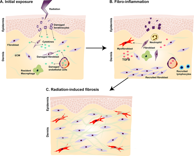
Radiotherapy (RT) has become an indispensable part of oncologic treatment protocols for a range of malignancies. However, a serious adverse effect of RT is radiodermatitis; almost 95% of patients develop moderate to severe skin reactions following radiation treatment. In the acute setting, these can be erythema, desquamation, ulceration, and pain. Chronically, soft tissue atrophy, alopecia, and stiffness can be noted. Radiodermatitis can delay oncologic treatment protocols and significantly impair quality of life. There is currently a paucity of effective treatment options and prevention strategies for radiodermatitis. Importantly, recent preclinical and clinical studies have suggested that fat grafting may be of therapeutic benefit, reversing detrimental changes to soft tissue following RT. This review outlines the damaging effects of RT on the skin and soft tissue as well as discusses available treatment options for radiodermatitis. Emerging strategies to mitigate detrimental, chronic radiation-induced changes are also presented.
Figures
References
-
- Mendelsohn FA, Divino CM, Reis ED & Kerstein MD Wound care after radiation therapy. Advances in skin & wound care 15, 216–224 (2002). - PubMed
-
- Delaney G, Jacob S, Featherstone C & Barton M The role of radiotherapy in cancer treatment: estimating optimal utilization from a review of evidence‐based clinical guidelines. Cancer: Interdisciplinary International Journal of the American Cancer Society 104, 1129–1137 (2005). - PubMed
-
- Porock D & Kristjanson L Skin reactions during radiotherapy for breast cancer: the use and impact of topical agents and dressings. European journal of cancer care 8, 143–153 (1999). - PubMed
-
- Porock D, Nikoletti S & Kristjanson L Management of radiation skin reactions: literature review and clinical application. Plastic Surgical Nursing 19, 185 (1999). - PubMed
-
- Martin M, Lefaix J-L & Delanian S TGF-β1 and radiation fibrosis: a master switch and a specific therapeutic target? International Journal of Radiation Oncology* Biology* Physics 47, 277–290 (2000). - PubMed
Publication types
MeSH terms
Grants and funding
LinkOut - more resources
Full Text Sources
Medical
