. 2020 Feb;145(2):679-697.e5.
doi: 10.1016/j.jaci.2019.08.029. Epub 2019 Sep 9.

Intrathymic adeno-associated virus gene transfer rapidly restores thymic function and long-term persistence of gene-corrected T cells

Affiliations

Intrathymic adeno-associated virus gene transfer rapidly restores thymic function and long-term persistence of gene-corrected T cells

Marie Pouzolles et al. J Allergy Clin Immunol. 2020 Feb.

Abstract

Background: Patients with T-cell immunodeficiencies are generally treated with allogeneic hematopoietic stem cell transplantation, but alternatives are needed for patients without matched donors. An innovative intrathymic gene therapy approach that directly targets the thymus might improve outcomes.

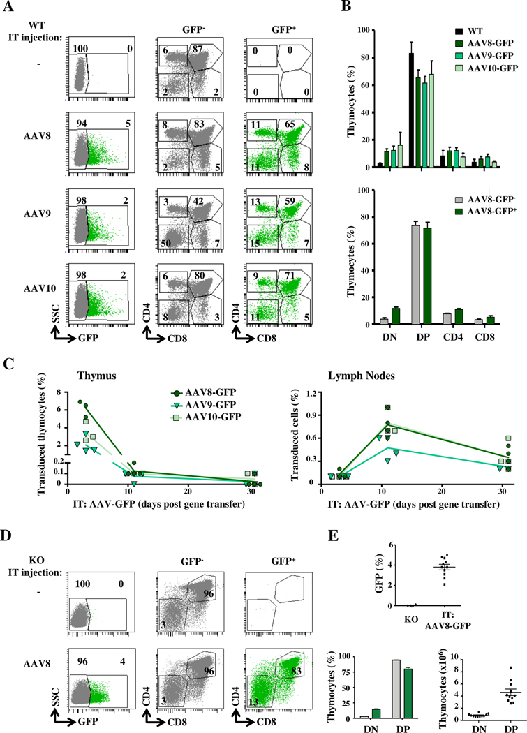
Objective: We sought to determine the efficacy of intrathymic adeno-associated virus (AAV) serotypes to transduce thymocyte subsets and correct the T-cell immunodeficiency in a zeta-associated protein of 70 kDa (ZAP-70)-deficient murine model.

Methods: AAV serotypes were injected intrathymically into wild-type mice, and gene transfer efficiency was monitored. ZAP-70-/- mice were intrathymically injected with an AAV8 vector harboring the ZAP70 gene. Thymus structure, immunophenotyping, T-cell receptor clonotypes, T-cell function, immune responses to transgenes and autoantibodies, vector copy number, and integration were evaluated.

Results: AAV8, AAV9, and AAV10 serotypes all transduced thymocyte subsets after in situ gene transfer, with transduction of up to 5% of cells. Intrathymic injection of an AAV8-ZAP-70 vector into ZAP-70-/- mice resulted in a rapid thymocyte differentiation associated with the development of a thymic medulla. Strikingly, medullary thymic epithelial cells expressing the autoimmune regulator were detected within 10 days of gene transfer, correlating with the presence of functional effector and regulatory T-cell subsets with diverse T-cell receptor clonotypes in the periphery. Although thymocyte reconstitution was transient, gene-corrected peripheral T cells harboring approximately 1 AAV genome per cell persisted for more than 40 weeks, and AAV vector integration was detected.

Conclusions: Intrathymic AAV-transduced progenitors promote a rapid restoration of the thymic architecture, with a single wave of thymopoiesis generating long-term peripheral T-cell function.

Keywords: Severe combined immunodeficiency; T-cell reconstitution; gene therapy; humoral immunity; intrathymic gene transfer; medulla formation; thymus; zeta-associated protein of 70kDa.


Figures

FIG 1.
FIG 1.. AAV 8, 9 and 10 serotypes efficiently transduce all thymocyte subsets.
A, scAAV 8, 9 and 10 serotypes harboring GFP were administered intrathymically (IT) into WT mice and GFP expression was assessed at day 3. Representative CD4/CD8 profiles from non-transduced (grey) and transduced (green) GFP+ thymocytes are presented. B, Quantification of the percentages of GFP+ double negative (DN), double positive (DP), single positive CD4 (SP4) and single positive CD8 (SP8) thymocytes are shown and compared to non-transduced WT mice (upper panel). Thymocyte subset repartition within non-transduced (GFP-) and transduced (GFP+) compartments of AAV8-GFP-transduced mice are presented (lower panel; means±SEM). C, The percentages of GFP+ thymocytes and lymph node (LN) T cells are presented as a function of time (n=3–5 mice per time point). D, Representative plots of GFP− and GFP+ thymocytes at day 3 following IT injection of the scAAV-GFP vector into ZAP-70−/− (KO) mice (left). Quantification of GFP+ cells in KO and AAV-transduced KO mice (n=11) are presented (top). The mean percentages of DN and DP thymocytes within the non-transduced (GFP−, grey) and transduced (GFP+, green) subsets (left bottom panel) as well as the absolute numbers of transduced GFP+ thymocytes are shown (right bottom panel).
FIG 2.
FIG 2.. Intrathymic AAV8-ZAP-70 gene transfer results in the rapid reconstitution of the thymic architecture in ZAP-70−/− mice.
A, ssAAV8-ZAP-70 or scAAV8-GFP virions were IT-injected into ZAP-70−/− (KO) mice and thymi from WT, KO and gene-transduced mice were evaluated for the presence of a thymic medulla by keratin14 (K14) staining and AIRE+ cells. Representative confocal images of thymic tissue sections at 1.5 (10 days), 3 and 10 weeks (w) post gene transfer are shown. B, The number of AIRE+ cells/mm2 of thymus were quantified in the indicated conditions. Each point represents the quantification of individual medulla derived from 3 distinct mice. C, Mean percentages of mature SP4 and SP8 thymocytes within the transduced GFP+ and ZAP-70+ subsets are shown in WT (left panel) and ZAP-70−/− (right panel) mice at the indicated time points following intrathymic AAV8-GFP and AAV8-ZAP-70 gene transfer, respectively. D, Absolute numbers of SP4 and SP8 thymocytes in WT mice (left panel) and AAV8-ZAP-70-transduced mice (right panel) were determined from the SP4 and SP8 gates at the indicated time points (n=5). Statistical significance was determined using an unpaired 2-tailed t-test or a one-way ANOVA with a Tukey’s multiple comparison test; *, p<0.05; **, p<0.01; ***, p<0.001; ****, p<0.0001.
FIG 3.
FIG 3.. Intrathymic AAV8 transduction results in long-term maintenance of peripheral T cells and T lineage-specific transgene expression.
A, The presence of peripheral blood T cells was monitored by flow cytometry following IT administration of AAV8-GFP or AAV8-ZAP-70 vectors and data from individual mice are presented at 3–43 weeks. Statistical differences between mice treated by IT AAV8-ZAP-70 gene transfer as compared to IT AAV8-GFP transfer or control KO mice were evaluated by a one-way ANOVA with a Tukey’s multiple comparison test for the following time groups; 3–10 weeks, 11–20 weeks, 21–30 weeks and 31–40 weeks (***, p<0.001, for all groups). B, The percentages of peripheral CD4+, CD8+, CD19+ and CD11b+ hematopoietic cells expressing ectopic ZAP-70 following IT AAV8-ZAP-70 transduction are presented. C, ZAP-70 protein expression in transduced T cells was evaluated by intracellular staining and MFI relative to WT T cells are presented at the indicated time points. D, Representative plots of CD3+ lymph node T cells are presented. Total T cell numbers (means±SEM) are shown (right). E, The repartition of CD4/CD8 lymphocytes and mean CD4/CD8 ratios are shown at the indicated time points. Statistical significance was determined by a 2-tailed unpaired t-test *, p<0.05.
FIG 4.
FIG 4.. AAV8-ZAP-70-transduced peripheral T lymphocytes exhibit stable vector genomes following TCR-induced proliferation and diverse TCR repertoires.
A, AAV genome copy number in lymph node samples was assessed by qPCR in IT AAV8-ZAP-70-transduced mice at the indicated time points. Vector genomes per diploid genome (Vg/Dg) were quantified relative to the albumin gene and normalized to the percentage of T cells. B, Proliferation was assessed following TCR stimulation by CTV fluorescence and representative histograms at day 0 (light grey) and day 4 (dark grey) are shown. AAV genome copy was monitored as above and quantifications (n=9) are shown (p, non-significant (ns)). C, Schematic representation of the AAV-ZAP-70 vector are shown. PCR products, representing integrated vector, are shown at the indicated time points and T cells from WT mice are presented as a negative control. D, The number of distinct TRA V, TRA J, TRA VJ, TRB V, TRB J, TRB VJ, TRA and TRB clonotypes are presented (top). Clonotype frequencies are presented with each clonotype represented by a dot whose size is proportional to its frequency. Violin plots represent the density of the distribution and black dots show the median frequency per repertoire (bottom).
FIG 5.
FIG 5.. Induction of a T cell-independent humoral response to AAV8 capsid epitopes following intrathymic vector administration.
A, Total IgG titers against OVA were monitored in non-immunized WT, KO and IT-AAV8-ZAP-70-treated KO mice (46w post gene transfer) and 6 weeks post immunization by ELISA. Reactivity was monitored as a function of dilution, as indicated (n=4). Statistical significance was evaluated at each dilution by a one-way ANOVA with a Tukey’s multiple comparison test for each dilution and is noted for significant differences; ***, p<0.001; **, p<0.01. B, Neutralizing antibodies (Nab) against the AAV8 serotype was monitored in WT and ZAP-70−/− mice following intrathymic AAV8-GFP and AAV8-ZAP-70 transduction. Serum dilutions of 1:10, 1:100 and 1:1000 are shown by the decreasing slope of the triangle, respectively, and Nab activity is defined as a decrease in AAV transduction of >50% compared to AAV incubation with PBS alone (red line). Each point represents serum from an individual mouse at 3, 10 and 17 weeks post transduction and mean levels are indicated by a horizontal line. C, Antibodies against ZAP-70 protein were measured by ELISA and data are presented as ng of antibody/ ml of serum. Antibody levels of >25ng/ml are considered significant.
FIG 6.
FIG 6.. Transient induction of a broad spectrum of antibodies in ZAP-70−/− mice following intrathymic AAV8 gene transfer remains significantly lower than that detected in autoimmune mice.
A, The presence of IgG autoantibodies in serum samples of ZAP-70−/− (KO) mice and following IT administration of either AAV8-GFP or AAV8-ZAP-70 vectors was evaluated at the indicated time points. IgG autoantibodies against 123 antigens were assessed by protein microarray and heat maps showing autoantibody reactivity relative to levels in age-matched WT mice are presented. B, The levels of auto-antibodies against RNP, dsDNA and SSA were evaluated by ELISA in control MRL/MpJ mice, autoimmune-prone MRL/lpr mice and IT:AAV8-ZAP-70-treated mice at 10 and 43 weeks post gene transfer. Each point represents serum from an individual mouse and statistical significance was determined by a one-way ANOVA with a Tukey’s multiple comparison test and significance is indicated; **, p<0.01; ***, p<0.001 ****, p<0.0001, ns-non significant.
FIG 7.
FIG 7.. AAV8-ZAP-70-transduced thymocytes differentiate into effector and regulatory T cells.
A, The presence of naïve (CD62L+CD44−, N), central memory (CD62L+ CD44+, CM), and effector memory (CD62L−CD44+, EM) cells within the CD4 and CD8 subsets were monitored at 3, 10, 17 and 43 weeks post IT-AAV8-ZAP-70 gene transfer. The percentages of cells in each subset were quantified at each time point and mean levels ± SEM (n=4–5 mice per time point) are presented. B, LN cells from the indicated mice were stimulated ex vivo with anti-CD3/anti-CD28 mAbs for 3 days and cytokine secretion was measured. Each point represents data from a single mouse and horizontal lines represent mean levels (pg/ml). C, The percentages of Foxp3+CD25+ peripheral Tregs was evaluated and representative dot plots are presented (left) together with mean percentages ± SEM at each time point (n=5–14 mice per time point, right). D, Conventional and regulatory CD4 T cell subsets were FACS-sorted on the basis of CD25 expression. Foxp3 and IL-10 transcripts in each subset were monitored by qRT-PCR (n=2 to 4 mice in 2 independent experiments). Statistical significance was determined by a 2-tailed unpaired t-test and significance is indicated; *, p<0.05; **, p<0.01; ***, p<0.001 ****, p<0.0001.

Comment in

Similar articles

Cited by

  • AAV integration in human hepatocytes.
    Dalwadi DA, Calabria A, Tiyaboonchai A, Posey J, Naugler WE, Montini E, Grompe M. Dalwadi DA, et al. Mol Ther. 2021 Oct 6;29(10):2898-2909. doi: 10.1016/j.ymthe.2021.08.031. Epub 2021 Aug 28. Mol Ther. 2021. PMID: 34461297 Free PMC article.
  • Lymphotoxin: from the physiology to the regeneration of the thymic function.
    Borelli A, Irla M. Borelli A, et al. Cell Death Differ. 2021 Aug;28(8):2305-2314. doi: 10.1038/s41418-021-00834-8. Epub 2021 Jul 22. Cell Death Differ. 2021. PMID: 34290396 Free PMC article. Review.
  • Delivering CRISPR to the HIV-1 reservoirs.
    Gurrola TE, Effah SN, Sariyer IK, Dampier W, Nonnemacher MR, Wigdahl B. Gurrola TE, et al. Front Microbiol. 2024 May 15;15:1393974. doi: 10.3389/fmicb.2024.1393974. eCollection 2024. Front Microbiol. 2024. PMID: 38812680 Free PMC article. Review.
  • Intrathymic AAV delivery results in therapeutic site-specific integration at TCR loci in mice.
    Calabria A, Cipriani C, Spinozzi G, Rudilosso L, Esposito S, Benedicenti F, Albertini A, Pouzolles M, Luoni M, Giannelli S, Broccoli V, Guilbaud M, Adjali O, Taylor N, Zimmermann VS, Montini E, Cesana D. Calabria A, et al. Blood. 2023 May 11;141(19):2316-2329. doi: 10.1182/blood.2022017378. Blood. 2023. PMID: 36790505 Free PMC article.
  • T-cell specific in vivo gene delivery with DART-AAVs targeted to CD8.
    Demircan MB, Zinser LJ, Michels A, Guaza-Lasheras M, John F, Gorol JM, Theuerkauf SA, Günther DM, Grimm D, Greten FR, Chlanda P, Thalheimer FB, Buchholz CJ. Demircan MB, et al. Mol Ther. 2024 Oct 2;32(10):3470-3484. doi: 10.1016/j.ymthe.2024.08.002. Epub 2024 Aug 8. Mol Ther. 2024. PMID: 39113357 Free PMC article.

References

    1. Al-Herz W, Bousfiha A, Casanova JL, Chatila T, Conley ME, Cunningham-Rundles C, et al. Primary immunodeficiency diseases: an update on the classification from the international union of immunological societies expert committee for primary immunodeficiency. Front Immunol. 2014;5:162. - PMC - PubMed
    1. Heimall J, Puck J, Buckley R, Fleisher TA, Gennery AR, Neven B, et al. Current Knowledge and Priorities for Future Research in Late Effects after Hematopoietic Stem Cell Transplantation (HCT) for Severe Combined Immunodeficiency Patients: A Consensus Statement from the Second Pediatric Blood and Marrow Transplant Consortium International Conference on Late Effects after Pediatric HCT. Biol Blood Marrow Transplant. 2017; 23(3):379–387. - PMC - PubMed
    1. Fischer A, Hacein-Bey-Abina S, Cavazzana-Calvo M. Gene therapy for primary immunodeficiencies. Hematology/oncology clinics of North America. 2011;25(1):89–100. - PubMed
    1. Aiuti A, Cattaneo F, Galimberti S, Benninghoff U, Cassani B, Callegaro L, et al. Gene therapy for immunodeficiency due to adenosine deaminase deficiency. N Engl J Med. 2009;360(5):447–58. - PubMed
    1. Aiuti A, Biasco L, Scaramuzza S, Ferrua F, Cicalese MP, Baricordi C, et al. Lentiviral hematopoietic stem cell gene therapy in patients with Wiskott-Aldrich syndrome. Science. 2013;341(6148):1233151. - PMC - PubMed

Publication types

Substances