. 2020 Jan;28(1):71-81.
doi: 10.1016/j.joca.2019.08.005. Epub 2019 Sep 9.

MRI-based screening for structural definition of eligibility in clinical DMOAD trials: Rapid OsteoArthritis MRI Eligibility Score (ROAMES)

Affiliations

MRI-based screening for structural definition of eligibility in clinical DMOAD trials: Rapid OsteoArthritis MRI Eligibility Score (ROAMES)

F W Roemer et al. Osteoarthritis Cartilage. 2020 Jan.

Abstract

Purpose: Our aim was to introduce a simplified MRI instrument, Rapid OsteoArthritis MRI Eligibility Score (ROAMES), for defining structural eligibility of patients for inclusion in disease-modifying osteoarthritis drug trials using a tri-compartmental anatomic approach that enables stratification of knees into different structural phenotypes and includes diagnoses of exclusion. We also aimed to define overlap between phenotypes and determine reliability.

Methods: 50 knees from the Foundation for National Institutes of Health Osteoarthritis Biomarkers study, a nested case-control study within the Osteoarthritis Initiative, were selected within pre-defined definitions of phenotypes as either inflammatory, subchondral bone, meniscus/cartilage, atrophic or hypertrophic. A focused scoring instrument was developed covering cartilage, meniscal damage, inflammation and osteophytes. Diagnoses of exclusion were meniscal root tears, osteonecrosis, subchondral insufficiency fracture, tumors, malignant marrow infiltration and acute traumatic changes. Reliability was determined using weighted kappa statistics. Descriptive statistics were used for determining concordance between the a priori phenotypic definition and ROAMES and overlap between phenotypes.

Results: ROAMES identified 43 of 50 (86%) pre-defined phenotypes correctly. Of the 50 participants, 27 (54%) had no additional phenotypes other than the pre-defined phenotype. 18 (36%) had one and 5 (10%) had two additional phenotypes. None had three or four additional phenotypes. All features of ROAMES showed almost perfect agreement. One case with osteonecrosis and one with a tumor were detected.

Conclusions: ROAMES is able to screen and stratify potentially eligible knees into different structural phenotypes and record relevant diagnoses of exclusion. Reliability of the instrument showed almost perfect agreement.

Keywords: Clinical trial; Eligibility; MRI; Osteoarthritis.


Conflict of interest statement

Conflict of interests

FWR is shareholder of Boston Imaging Core Lab. (BICL),LLC.

CKK is consultant to EMD Serono, Thusane, Express Scripts, Regulus, GSK, Regeneron, Fidia, Taiwan Liposome Company.

TN is consultant to Pfizer, EMD Merck Serono, Novartis.

DJH is consultant to Merck Serono, Pfizer, Tissuegene, TLCBio.

MH received an institutional grant by the NIH and is consultant to EMD Serono.

JEC is consultant to Boston Imaging Core Lab (BICL),LLC.

AG has received consultancies, speaking fees, and/or honoraria from Sanofi-Aventis, Merck Serono, and TissuGene and is President and shareholder of Boston Imaging Core Lab (BICL), LLC a company providing image assessment services.

DTF and JL have no conflict to declare.

Figures

Figure 1.
Figure 1.
Diagnoses of Exclusion: A. Posterior meniscal root tears are defined by avulsion of the meniscal ligamentous root (white arrow) or by complete radial tears close to the tibial attachment of the meniscus (black arrow). These tears result in biomechanical alterations comparable to a complete meniscectomy and may lead to rapid cartilage loss and potentially joint collapse. B. Coronal intermediate-weighted fat-suppressed image shows a medial posterior root tear (arrow). C. Subchondral insufficiency fractures are defined by a hypointense fracture line adjacent to the subchondral plate surrounded by a large area of bone marrow edema. Subchondral insufficiency fractures may heal or may progress to articular osteochondral defects and joint collapse. D. Coronal intermediate-weighted image depicts a subchondral insufficiency fracture (arrow) with a large area of surrounding bone marrow edema. E. Bone infarct or avascular necrosis is defined by a serpiginous area of hyperintensity on fluid sensitive fat-suppressed sequences and a fat-equivalent center. These are commonly observed in conjunction with systemic disease or steroid therapy and may increase the risk of joint collapse particularly when in an epiphyseal subchondral location. F. A large area of epiphyseal osteonecrosis is shown on this sagittal intermediate weighted fat-suppressed image. Arrows point to the demarcation line. Infarcts are characterized by a serpiginous hyperintense demarcation and a fat-equivalent center. In addition, there is an area of cartilage delamination at the posterior lateral femur (short arrow). This fragment is at high risk of detachment and developing into an osteochondral defect and subsequent possible joint collapse. G. Diffuse cellular bone marrow infiltration due to malignancy such as lymphoma or leukemia may be diagnosed by MRI and needs to be differentiated from red marrow reconversion in anemia. The latter is not observed in an epiphyseal location while cellular infiltration is. H. Sagittal intermediate-weighted fat-suppressed image shows diffuse marrow infiltration due to acute lymphatic leukemia. Cellular infiltrations characterized by diffuse hyperintensity on fluid-sensitive sequences including the epiphyses as well as metaphyses. I. Pigmented villo-nodular synovitis (PVNS) or diffuse-type giant cell tumor of the tendon sheath (dGCTTS) are solid, benign tumors of the articular cavity that need surgical treatment and tend to recur. These are commonly observed adjacent to Hoffa’s fat pad in the anterior knee compartment as shown here. J. Sagittal intermediate-weighted image shows an oval-shaped lesion of intermediate signal intensity (arrows) in the anterior compartment of the knee joint. K. Coronal intermediate-weighted image shows the same lesion in the lateral gutter of the patello-femoral joint (short arrows). Long arrow points to focal intralesional hypointensities characteristic of representing hemosiderin deposits. L. Acute articular damage may result in a spectrum of morphologic findings from subchondral contusions to chondral flake fractures or osteochondral depression, as shown in this example. Commonly traumatic bone marrow edema (also called contusion) is seen in a subchondral location (arrows). In addition, there is a sharply delineated chondral depression (arrowheads) and also a contour deformity of the subchondral plate in this illustration. M. Sagittal intermediate-weighted fat-suppressed image shows a large bone contusion (i.e. traumatic bone marrow lesion) in the posterior lateral tibia. In addition, there is an osteochondral depression with disruption of the subchondral plate (arrow).”
Figure 1.
Figure 1.
Diagnoses of Exclusion: A. Posterior meniscal root tears are defined by avulsion of the meniscal ligamentous root (white arrow) or by complete radial tears close to the tibial attachment of the meniscus (black arrow). These tears result in biomechanical alterations comparable to a complete meniscectomy and may lead to rapid cartilage loss and potentially joint collapse. B. Coronal intermediate-weighted fat-suppressed image shows a medial posterior root tear (arrow). C. Subchondral insufficiency fractures are defined by a hypointense fracture line adjacent to the subchondral plate surrounded by a large area of bone marrow edema. Subchondral insufficiency fractures may heal or may progress to articular osteochondral defects and joint collapse. D. Coronal intermediate-weighted image depicts a subchondral insufficiency fracture (arrow) with a large area of surrounding bone marrow edema. E. Bone infarct or avascular necrosis is defined by a serpiginous area of hyperintensity on fluid sensitive fat-suppressed sequences and a fat-equivalent center. These are commonly observed in conjunction with systemic disease or steroid therapy and may increase the risk of joint collapse particularly when in an epiphyseal subchondral location. F. A large area of epiphyseal osteonecrosis is shown on this sagittal intermediate weighted fat-suppressed image. Arrows point to the demarcation line. Infarcts are characterized by a serpiginous hyperintense demarcation and a fat-equivalent center. In addition, there is an area of cartilage delamination at the posterior lateral femur (short arrow). This fragment is at high risk of detachment and developing into an osteochondral defect and subsequent possible joint collapse. G. Diffuse cellular bone marrow infiltration due to malignancy such as lymphoma or leukemia may be diagnosed by MRI and needs to be differentiated from red marrow reconversion in anemia. The latter is not observed in an epiphyseal location while cellular infiltration is. H. Sagittal intermediate-weighted fat-suppressed image shows diffuse marrow infiltration due to acute lymphatic leukemia. Cellular infiltrations characterized by diffuse hyperintensity on fluid-sensitive sequences including the epiphyses as well as metaphyses. I. Pigmented villo-nodular synovitis (PVNS) or diffuse-type giant cell tumor of the tendon sheath (dGCTTS) are solid, benign tumors of the articular cavity that need surgical treatment and tend to recur. These are commonly observed adjacent to Hoffa’s fat pad in the anterior knee compartment as shown here. J. Sagittal intermediate-weighted image shows an oval-shaped lesion of intermediate signal intensity (arrows) in the anterior compartment of the knee joint. K. Coronal intermediate-weighted image shows the same lesion in the lateral gutter of the patello-femoral joint (short arrows). Long arrow points to focal intralesional hypointensities characteristic of representing hemosiderin deposits. L. Acute articular damage may result in a spectrum of morphologic findings from subchondral contusions to chondral flake fractures or osteochondral depression, as shown in this example. Commonly traumatic bone marrow edema (also called contusion) is seen in a subchondral location (arrows). In addition, there is a sharply delineated chondral depression (arrowheads) and also a contour deformity of the subchondral plate in this illustration. M. Sagittal intermediate-weighted fat-suppressed image shows a large bone contusion (i.e. traumatic bone marrow lesion) in the posterior lateral tibia. In addition, there is an osteochondral depression with disruption of the subchondral plate (arrow).”
Figure 2.
Figure 2.
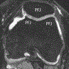
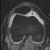
Compartmental scoring approach in ROAMES. While subregions are defined in identical fashion as in MOAKS, only maximum grades per compartment are recorded. Only the maximum subregional scores of the patello-femoral, the medial and lateral tibio-femoral joints are considered. Figure depicts MOAKS subregions relabeled as compartmental categories as defined in ROAMES. A. Patello-femoral joint. The four MOAKS subregions of medial and lateral patella and the medial and lateral femoral trochlea are combined as patello-femoral joint. B. Sagittal intermediate-weighted fat suppressed image shows MOAKS subregions and assignment to the lateral tibio-femoral and the patello-femoral joints. C Coronal intermediate-weighted image shows the medial and lateral tibio-femoral compartments. Note that identical to MOAKS the femoral notch is part of the medial tibio-femoral joint.
Figure 2.
Figure 2.
Compartmental scoring approach in ROAMES. While subregions are defined in identical fashion as in MOAKS, only maximum grades per compartment are recorded. Only the maximum subregional scores of the patello-femoral, the medial and lateral tibio-femoral joints are considered. Figure depicts MOAKS subregions relabeled as compartmental categories as defined in ROAMES. A. Patello-femoral joint. The four MOAKS subregions of medial and lateral patella and the medial and lateral femoral trochlea are combined as patello-femoral joint. B. Sagittal intermediate-weighted fat suppressed image shows MOAKS subregions and assignment to the lateral tibio-femoral and the patello-femoral joints. C Coronal intermediate-weighted image shows the medial and lateral tibio-femoral compartments. Note that identical to MOAKS the femoral notch is part of the medial tibio-femoral joint.
Figure 3.
Figure 3.
Phenotypic characterization. A. The inflammatory phenotype is characterized by large joint effusion (asterisk) and so-called Hoffa-synovitis, a non-specific surrogate of whole knee synovitis. B. Bone phenotype. A large bone marrow lesion (BML) is present in the medial central subregion of the medial femur (grade 3, arrows). The maximum grade 3 BML defines this knee as a bone phenotype. C. The cartilage/meniscus phenotype is characterized by severe meniscal damage depicted in this example as partial meniscal maceration of the medial meniscal body (arrowhead) and commonly associated with severe cartilage loss (arrows point to diffuse superficial cartilage damage of the medial femur). In addition there is diffuse superficial cartilage damage at the medial tibia. D. The atrophic phenotype is characterized by severe cartilage loss without relevant osteophyte formation. There is marked cartilage damage in the medial compartment (arrows) and meniscal maceration (arrowhead) but no relevant osteophyte formation. This knee also fulfilled definition of the cartilage/meniscus phenotype. E. The hypertrophic phenotype is characterized by large osteophytes with only minimal cartilage loss and is defined commonly in a compartmental manner. The arrow points to a grade 3 osteophyte according to ROAMES and a grade 2 osteophyte is depicted by the arrowhead at the medial tibia.
Figure 3.
Figure 3.
Phenotypic characterization. A. The inflammatory phenotype is characterized by large joint effusion (asterisk) and so-called Hoffa-synovitis, a non-specific surrogate of whole knee synovitis. B. Bone phenotype. A large bone marrow lesion (BML) is present in the medial central subregion of the medial femur (grade 3, arrows). The maximum grade 3 BML defines this knee as a bone phenotype. C. The cartilage/meniscus phenotype is characterized by severe meniscal damage depicted in this example as partial meniscal maceration of the medial meniscal body (arrowhead) and commonly associated with severe cartilage loss (arrows point to diffuse superficial cartilage damage of the medial femur). In addition there is diffuse superficial cartilage damage at the medial tibia. D. The atrophic phenotype is characterized by severe cartilage loss without relevant osteophyte formation. There is marked cartilage damage in the medial compartment (arrows) and meniscal maceration (arrowhead) but no relevant osteophyte formation. This knee also fulfilled definition of the cartilage/meniscus phenotype. E. The hypertrophic phenotype is characterized by large osteophytes with only minimal cartilage loss and is defined commonly in a compartmental manner. The arrow points to a grade 3 osteophyte according to ROAMES and a grade 2 osteophyte is depicted by the arrowhead at the medial tibia.

References

    1. Hunter DJ, Schofield D, Callander E. The individual and socioeconomic impact of osteoarthritis. Nat Rev Rheumatol 2014;10:437–41. - PubMed
    1. Karsdal MA, Byrjalsen I, Henriksen K, Riis BJ, Lau EM, Arnold M, et al. The effect of oral salmon calcitonin delivered with 5-CNAC on bone and cartilage degradation in osteoarthritic patients: a 14-day randomized study. Osteoarthritis Cartilage 2010;18:150–9. - PubMed
    1. Spector TD, Conaghan PG, Buckland-Wright JC, Garnero P, Cline GA, Beary JF, et al. Effect of risedronate on joint structure and symptoms of knee osteoarthritis: results of the BRISK randomized, controlled trial [ISRCTN01928173]. Arthritis Res Ther 2005;7:R625–33. - PMC - PubMed
    1. Alexandersen P, Karsdal MA, Byrjalsen I, Christiansen C. Strontium ranelate effect in postmenopausal women with different clinical levels of osteoarthritis. Climacteric 2011;14:236–43. - PubMed
    1. Reginster JY, Badurski J, Bellamy N, Bensen W, Chapurlat R, Chevalier X, et al. Efficacy and safety of strontium ranelate in the treatment of knee osteoarthritis: results of a double-blind, randomised placebo-controlled trial. Ann Rheum Dis 2013;72:179–86. - PMC - PubMed

Publication types