. 2019 Sep 11;11(9):1345.
doi: 10.3390/cancers11091345.

Targeting Autophagy by MPT0L145, a Highly Potent PIK3C3 Inhibitor, Provides Synergistic Interaction to Targeted or Chemotherapeutic Agents in Cancer Cells

Affiliations

Targeting Autophagy by MPT0L145, a Highly Potent PIK3C3 Inhibitor, Provides Synergistic Interaction to Targeted or Chemotherapeutic Agents in Cancer Cells

Chun-Han Chen et al. Cancers (Basel). .

Abstract

Anticancer therapies reportedly promote pro-survival autophagy in cancer cells that confers drug resistance, rationalizing the concept to combine autophagy inhibitors to increase their therapeutic potential. We previously identified that MPT0L145 is a PIK3C3/FGFR inhibitor that not only increases autophagosome formation due to fibroblast growth factor receptor (FGFR) inhibition but also perturbs autophagic flux via PIK3C3 inhibition in bladder cancer cells harboring FGFR activation. In this study, we hypothesized that combined-use of MPT0L145 with agents that induce pro-survival autophagy may provide synthetic lethality in cancer cells without FGFR activation. The results showed that MPT0L145 synergistically sensitizes anticancer effects of gefitinib and gemcitabine in non-small cell lung cancer A549 cells and pancreatic cancer PANC-1 cells, respectively. Mechanistically, drug combination increased incomplete autophagy due to impaired PIK3C3 function by MPT0L145 as evidenced by p62 accumulation and no additional apoptotic cell death was observed. Meanwhile, drug combination perturbed survival pathways and increased vacuolization and ROS production in cancer cells. In conclusion, the data suggest that halting pro-survival autophagy by targeting PIK3C3 with MPT0L145 significantly sensitizes cancer cells to targeted or chemotherapeutic agents, fostering rational combination strategies for cancer therapy in the future.

Keywords: PIK3C3; autophagy; gefitinib; gemcitabine; synergism.


Conflict of interest statement

The authors declare no conflict of interest.

Figures

Figure 1
Figure 1
Effects of targeted or chemotherapeutic agents on autophagy in cancer cells. (A) A549 and (B) PANC-1 cells were treated with indicated drugs for 24h and subjected to western blot analysis by antibodies against LC3B and GAPDH.
Figure 2
Figure 2
MPT0L145 sensitized cancer cells to targeted or chemotherapeutic agents. (A,C) A549 cells were treated with indicated concentrations of gefitinib in the absence or presence of MPT0L145 for 72h and subjected to MTT assay (A) or trypan blue exclusion assay (C). Data are expressed as means ± S.D. (N = 3, * p < 0.05, *** p < 0.001 compared to gefitinib alone). (B,D) PANC-1 cells were treated with indicated concentrations of gemcitabine in the absence or presence of MPT0L145 for 72h and subjected to MTT assay (B) or trypan blue exclusion assay (D). Data are expressed as means ± S.D. (N = 3, * p < 0.05, ** p < 0.01, *** p < 0.001 compared to gemcitabine alone).
Figure 3
Figure 3
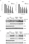
Effects of drug combination or PIK3C3-knockdown on autophagy in cancer cells. (A) PIK3C3 was stably knocked down in A549 (left panel) and PANC-1 cells (right panel) and exposed to indicated concentrations of gefitinib and gemcitabine, respectively. Cell viability of parental or PIK3C3-knockdown cells were measured by MTT assay. Data are expressed as means ± S.D. (N = 3, ** p < 0.01, *** p < 0.001 compared to wild-type group). (B) A549 cells were treated with gefitinib in the presence of MPT0L145 in parental cells or gefitinib alone in PIK3C3-knockdown cells for 24h and subjected to western blot analysis. (C) PANC-1 cells were treated with gemcitabine in the presence of MPT0L145 in parental cells or gemcitabine alone in PIK3C3-knockdown cells for 24h and subjected to western blot analysis.
Figure 4
Figure 4
Effects of drug combination on cell cycle distribution and apoptosis. (A,C) A549 and (B,D) PANC-1 cells were respectively treated with MPT0L145 (4 μM) in the presence of gefitinib or gemcitabine for 72h. The cells were then stained with propidium iodide solution (A,B) or Annexin V-FITC/PI solution (C,D) and analyzed by flow cytometry. Paclitaxel (Taxol, 0.1 μM) was included as a positive control of apoptosis. (E) A549 and (F) PANC-1 cells were exposed to MPT0L145 (4 μM) in the presence or absence of gefitinib or gemcitabine, respectively for 72h. The cells were subjected to western blot analysis by using antibodies against PARP, caspase-3 and GAPDH.
Figure 5
Figure 5
Effects of drug combination on cell survival pathways in cancer cells. (A) A549 and (B) PANC-1 cells were treated with MPT0L145 (L145, 4 μM) in combination with gefitinib or gemcitabine, respectively for 72h. The cells were then subjected to western blot analysis.
Figure 6
Figure 6
Drug combination increases intracellular vacuolization and reactive oxygen species (ROS) level in cancer cells. (A) A549 (upper panel) and PANC-1 (lower panel) cells were exposed to MPT0L145 (4 μM) alone and in combination with gefitinib (10 μM) or gemcitabine (100 μM) for 48h, respectively. Cell morphology was examined with the EVOS XL Core Cell Imaging System (Thermo Scientific). Intracellular vacuoles are indicated by white arrowheads. Scale bars: 100 μm. (B) A549 and (C) PANC-1 cells were treated with MPT0L145 (L145, 4 μM) in the presence of gefitinib (Gef, 10 μM) or gemcitabine (Gem, 100 μM), respectively for 72h. The cells were stained with H2DCFDA and intracellular ROS levels were measured by flow cytometry. Data are expressed as means ± S.D. (N = 3, * p < 0.05, ** p < 0.01 compared to control group; # p < 0.05, ## p < 0.01, ### p < 0.001 compared to combination group). (D) A549 and (E) PANC-1 cells were treated with MPT0L145 (L145, 4 μM) in the presence of gefitinib (Gef, 10 μM) or gemcitabine (Gem, 100 μM), respectively for 72h. Culture media or total cell lysates (TCL) from untreated cells, which served as maximum LDH release control, were collected and subjected to LDH analysis. Data were represented by the percentage of control group.

References

    1. Kaur J., Debnath J. Autophagy at the crossroads of catabolism and anabolism. Nat. Rev. Mol. Cell Biol. 2015;16:461–472. doi: 10.1038/nrm4024. - DOI - PubMed
    1. Boya P., Reggiori F., Codogno P. Emerging regulation and functions of autophagy. Nat. Cell Biol. 2013;15:713–720. doi: 10.1038/ncb2788. - DOI - PMC - PubMed
    1. Yang J., Carra S., Zhu W.G., Kampinga H.H. The regulation of the autophagic network and its implications for human disease. Int. J. Biol. Sci. 2013;9:1121–1133. doi: 10.7150/ijbs.6666. - DOI - PMC - PubMed
    1. Kumar A., Singh U.K., Chaudhary A. Targeting autophagy to overcome drug resistance in cancer therapy. Future Med. Chem. 2015;7:1535–1542. doi: 10.4155/fmc.15.88. - DOI - PubMed
    1. Sui X., Chen R., Wang Z., Huang Z., Kong N., Zhang M., Han W., Lou F., Yang J., Zhang Q., et al. Autophagy and chemotherapy resistance: a promising therapeutic target for cancer treatment. Cell Death Dis. 2013;4:e838. doi: 10.1038/cddis.2013.350. - DOI - PMC - PubMed

LinkOut - more resources