

Radiomic signatures with contrast-enhanced magnetic resonance imaging for the assessment of breast cancer receptor status and molecular subtypes: initial results
- PMID: 31514736
- PMCID: PMC6739929
- DOI: 10.1186/s13058-019-1187-z
Radiomic signatures with contrast-enhanced magnetic resonance imaging for the assessment of breast cancer receptor status and molecular subtypes: initial results
Abstract
Background: To evaluate the diagnostic performance of radiomic signatures extracted from contrast-enhanced magnetic resonance imaging (CE-MRI) for the assessment of breast cancer receptor status and molecular subtypes.
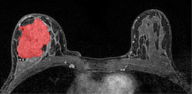
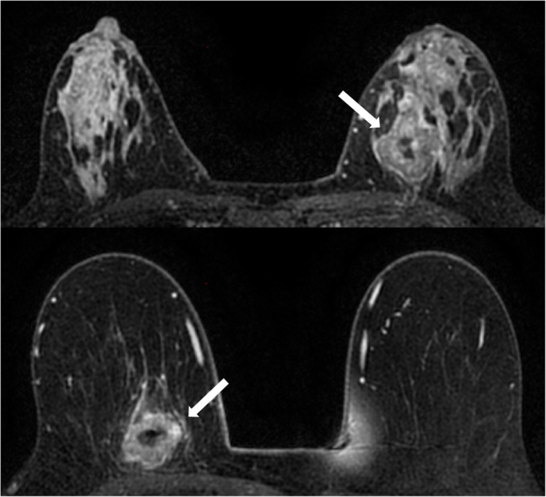
Methods: One hundred and forty-three patients with biopsy-proven breast cancer who underwent CE-MRI at 3 T were included in this IRB-approved HIPAA-compliant retrospective study. The training dataset comprised 91 patients (luminal A, n = 49; luminal B, n = 8; HER2-enriched, n = 11; triple negative, n = 23), while the validation dataset comprised 52 patients from a second institution (luminal A, n = 17; luminal B, n = 17; triple negative, n = 18). Radiomic analysis of manually segmented tumors included calculation of features derived from the first-order histogram (HIS), co-occurrence matrix (COM), run-length matrix (RLM), absolute gradient (GRA), autoregressive model (ARM), discrete Haar wavelet transform (WAV), and lesion geometry (GEO). Fisher, probability of error and average correlation (POE + ACC), and mutual information coefficients were used for feature selection. Linear discriminant analysis followed by k-nearest neighbor classification (with leave-one-out cross-validation) was used for pairwise radiomic-based separation of receptor status and molecular subtypes. Histopathology served as the standard of reference.
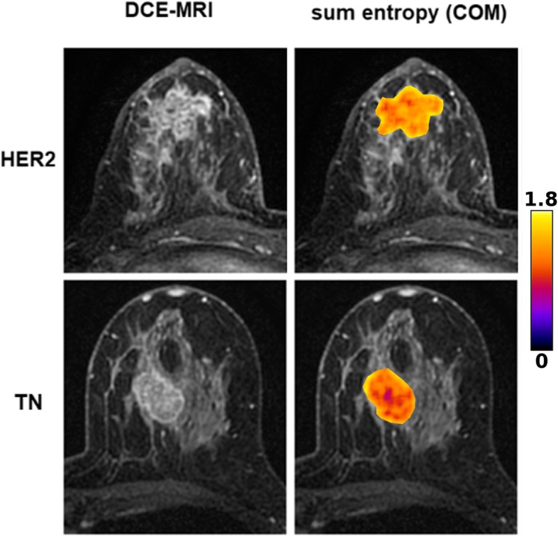
Results: In the training dataset, radiomic signatures yielded the following accuracies > 80%: luminal B vs. luminal A, 84.2% (mainly based on COM features); luminal B vs. triple negative, 83.9% (mainly based on GEO features); luminal B vs. all others, 89% (mainly based on COM features); and HER2-enriched vs. all others, 81.3% (mainly based on COM features). Radiomic signatures were successfully validated in the separate validation dataset for luminal A vs. luminal B (79.4%) and luminal B vs. triple negative (77.1%).
Conclusions: In this preliminary study, radiomic signatures with CE-MRI enable the assessment of breast cancer receptor status and molecular subtypes with high diagnostic accuracy. These results need to be confirmed in future larger studies.
Keywords: Breast cancer; Contrast-enhanced; Magnetic resonance imaging; Molecular subtype; Radiomics.
Conflict of interest statement
Elizabeth A. Morris and Katja Pinker were in part funded by the project: Use Of ctNA To Distinguish Between Benign and Malignant BIRADS 4 Radiographic Lesions. Maxine S Jochelson has received speaker honoraria from GE Healthcare. The other authors declare that they have no competing interests.
Figures
References
Publication types
MeSH terms
Substances
Grants and funding
LinkOut - more resources
Full Text Sources
Medical
Research Materials
Miscellaneous
