. 2019 Dec 12;54(6):1900378.
doi: 10.1183/13993003.00378-2019. Print 2019 Dec.

Suppression of HIF2 signalling attenuates the initiation of hypoxia-induced pulmonary hypertension

Affiliations

Suppression of HIF2 signalling attenuates the initiation of hypoxia-induced pulmonary hypertension

Cheng-Jun Hu et al. Eur Respir J. .

Abstract

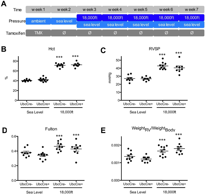
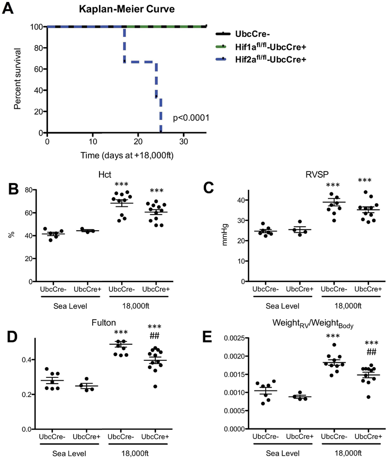
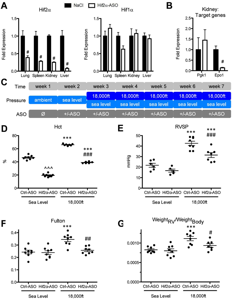
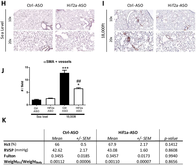
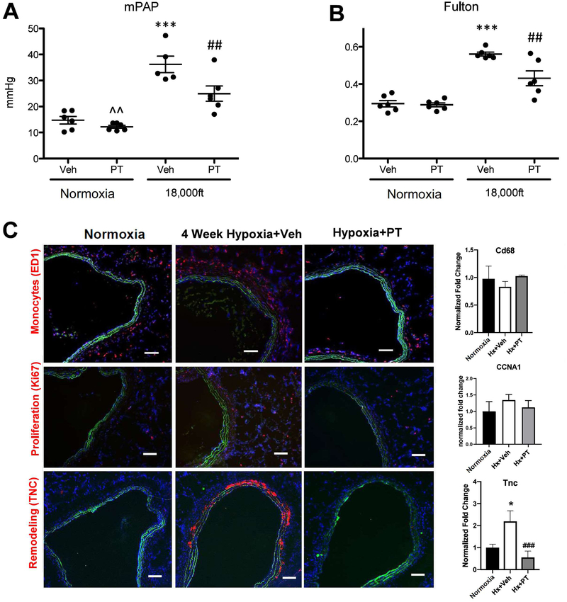
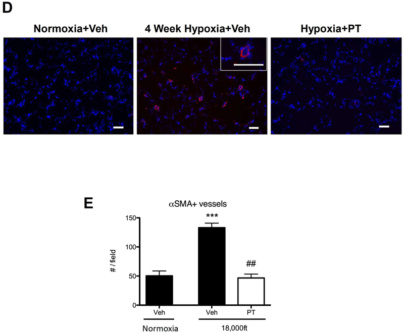
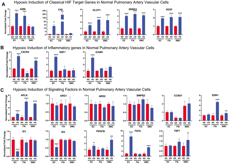
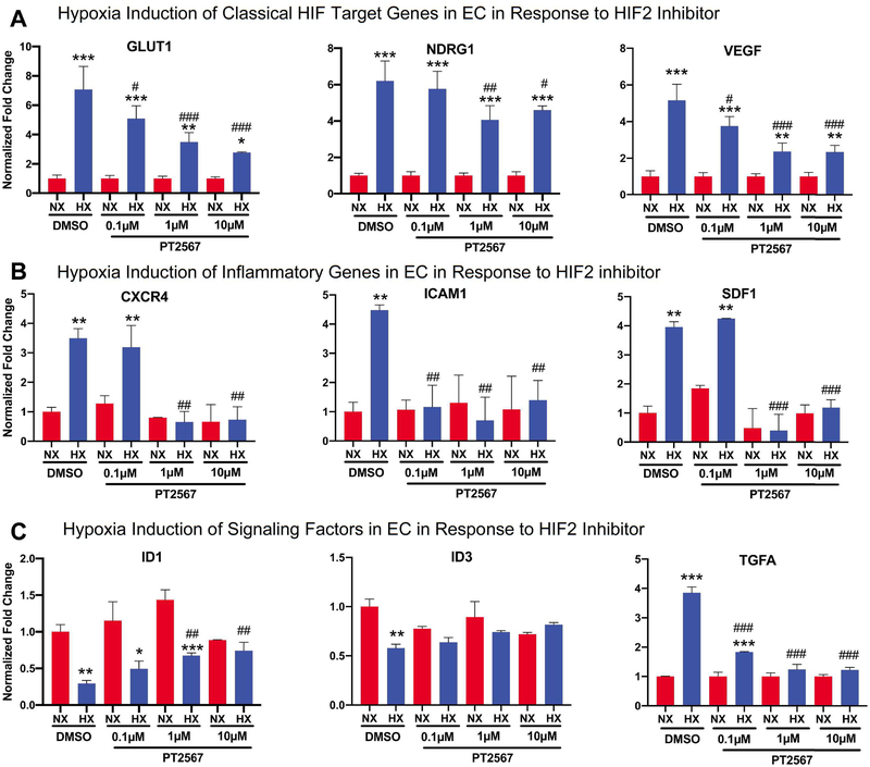
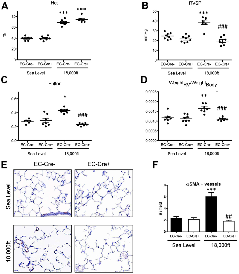
Most published studies addressing the role of hypoxia inducible factors (HIFs) in hypoxia-induced pulmonary hypertension development employ models that may not recapitulate the clinical setting, including the use of animals with pre-existing lung/vascular defects secondary to embryonic HIF ablation or activation. Furthermore, critical questions including how and when HIF signalling contributes to hypoxia-induced pulmonary hypertension remain unanswered.Normal adult rodents in which global HIF1 or HIF2 was inhibited by inducible gene deletion or pharmacological inhibition (antisense oligonucleotides (ASO) and small molecule inhibitors) were exposed to short-term (4 days) or chronic (4-5 weeks) hypoxia. Haemodynamic studies were performed, the animals euthanised, and lungs and hearts obtained for pathological and transcriptomic analysis. Cell-type-specific HIF signals for pulmonary hypertension initiation were determined in normal pulmonary vascular cells in vitro and in mice (using cell-type-specific HIF deletion).Global Hif1a deletion in mice did not prevent hypoxia-induced pulmonary hypertension at 5 weeks. Mice with global Hif2a deletion did not survive long-term hypoxia. Partial Hif2a deletion or Hif2-ASO (but not Hif1-ASO) reduced vessel muscularisation, increases in pulmonary arterial pressures and right ventricular hypertrophy in mice exposed to 4-5 weeks of hypoxia. A small molecule HIF2 inhibitor (PT2567) significantly attenuated early events (monocyte recruitment and vascular cell proliferation) in rats exposed to 4 days of hypoxia, as well as vessel muscularisation, tenascin C accumulation and pulmonary hypertension development in rats exposed to 5 weeks of hypoxia. In vitro, HIF2 induced a distinct set of genes in normal human pulmonary vascular endothelial cells, mediating inflammation and proliferation of endothelial cells and smooth muscle cells. Endothelial Hif2a knockout prevented hypoxia-induced pulmonary hypertension in mice.Inhibition of HIF2 (but not HIF1) can provide a therapeutic approach to prevent the development of hypoxia-induced pulmonary hypertension. Future studies are needed to investigate the role of HIFs in pulmonary hypertension progression and reversal.


Conflict of interest statement

Conflict of interest: C-J. Hu has nothing to disclose. Conflict of interest: J.M. Poth has nothing to disclose. Conflict of interest: H. Zhang has nothing to disclose. Conflict of interest: A. Flockton has nothing to disclose. Conflict of interest: A. Laux has nothing to disclose. Conflict of interest: S. Kumar has nothing to disclose. Conflict of interest: B. McKeon has nothing to disclose. Conflict of interest: G. Mouradian has nothing to disclose. Conflict of interest: M. Li has nothing to disclose. Conflict of interest: S. Riddle has nothing to disclose. Conflict of interest: S.C. Pugliese has nothing to disclose. Conflict of interest: R.D. Brown has nothing to disclose. Conflict of interest: E.M. Wallace has a patent “Compositions for Use in Treating Pulmonary Arterial Hypertension” pending to Peloton Therapeutics. Conflict of interest: B.B. Graham reports grants from National Institutes of Health, during the conduct of the study. Conflict of interest: M.G. Frid has nothing to disclose. Conflict of interest: K.R. Stenmark reports personal fees for advisory board or steering committee work from Pfizer, New York, Actelion (Entelligence) and Janssen Research and Development, outside the submitted work.

Figures

Figure 1:
Figure 1:. Global Hif1α is dispensable in establishing hypoxia-induced PH at 5 weeks in adult mice.
A) Experimental setup: Tamoxifen was injected into mice daily during week 1 to activate Cre and delete Hif1α gene, then all mice were moved to sea levels for one week, followed by exposing mice to either normoxia (PB-740mmHg) or hypoxia (simulated altitude of 18,000ft, PB=370 mmHg) for 5 weeks. After five weeks hypoxia exposure, readout parameters were measured and animals were euthanized. B) Hematocrit (Hct) levels. C) Right-ventricular systolic pressure (RVSP). D) Fulton index (ratio of weight of the RV to the weight of the LV and septum) and E) Ratio of RV-weight to bodyweight. Animal number in each group, under normoxia or hypoxia can be found in Fulton Index panel (Fig 1D). Due to technical difficulties, we were not able to obtain readings of Hct and/or RVSP for some mice, thus the animal number for these data was typically less than the animal number for Fulton index in this and other Figures of this study. * is for difference between hypoxia versus normoxia in the same genotype (or treatment) group in this and all other figures in the paper. Statistical analysis showed here is 2-way ANOVA analysis.
Figure 2:
Figure 2:. Global Hif2α gene deletion is incompatible for mouse survival under hypoxic conditions while global partial Hif2α gene deletion diminishes hypoxia-induced PH development at 5 weeks in adult mice.
Mice were treated with tamoxifen and exposed to normoxia or hypoxia as described in Fig 1A. A) Kaplan-Meier-Curve for survival of Hif2αfl/fl;UBC-CreERT+ mice during hypoxia exposure, compared to survival of Hif1αfl/fl;UBC-CreERT+ and Hif2αfl/fl;UBC-CreERT mice during exposure to hypoxia. More than 9 mice in each group were used for this experiment. B–E: Hemodynamics of Hif2αfl/WT;UbcCreERT+ and Hif2αfl/WT;UbcCreER mice after 5 week exposure to normoxia or hypoxia. B) Hematocrit (Hct) levels. C) RVSP. D) Fulton index. E) ratio of RV-weight to bodyweight. # is used to show the differences between genotypes or treatments under hypoxic condition in this and all other figures of this study. Statistical analysis showed here is 2-way ANOVA analysis (note in panel C there is a significant decrease in RVSP at 18,000 in UbcCre+ animals analyzed by unpaired T-test p<0.05).
Figure 3:
Figure 3:. Knockdown of Hif2α but not Hif1α utilizing antisense-oligonucleotides significantly reduces development of hypoxia-induced PH at 5 weeks in adult mice.
A and B) Testing the effectiveness and specificity of antisense-oligonucleotide in a pilot experiment. Wild-type C57bl/6J mice were treated either with injections of an antisense-oligonucleotide targeting Hif2α mRNA (Hif2α-ASO) or equal volumes of 0.9% NaCl (Ctrl) at days 1, 4, 8 and 11. At day 12, mice (N=3) were sacrificed and multiple organs were collected for RNA preparation. A) Levels of Hif1α and of Hif2α mRNA were quantified by qRT-PCR in indicated organs. B) Levels of Pgk1 (a HIF1 target gene) and Epo (a HIF2 target gene) in kidneys from mice targeted with NaCL or Hif2α-ASO, were quantified by qRT-PCR. C) Experimental setup. In week 2, mice were kept under sea level and began to receiving injection of Control, Hif1α-ASO, or Hif2α-ASO (two injections per week at Monday and Thursday). Starting week three, mice were exposed to either sea level or 18,000ft for 5 weeks, in which two injections per week were maintained. D–K) Endpoint measurements for the experimental animals in C. D) Hct. E) RVSP. F) Fulton index. G) ratio of RV-weight to bodyweight. H–J) alpha-SM-actin positive pulmonary vessels. K) Summary of Hct, RVSP, Fulton index, ratio of RV-weight to bodyweight for mice targeted with control or Hif1α-ASO under normoxia or hypoxia. Statistical significance as determined by t-test (A, B, J, and K) or 2-way ANOVA (D–G). ^ is used to show the differences between genotypes or treatments under normoxic condition in this and all other figures of this study.
Figure 4:
Figure 4:. Small molecule Hif2 inhibitor PT2567 significantly attenuates development of hypoxia-induced PH at 4 weeks in adult rats.
Sprague Dawley (SD) male rats weighing 210–245 grams (Charles River Laboratories) were housed in chambers under normoxia or hypoxic (high altitude ~ 18,000 feet) conditions for four weeks. Rats were dosed with vehicle methylcellulose (0.5%)/Tween-80 (0.5%) or PT2567 (300 mg/kg/day), beginning the day they were placed in chambers. After 4 weeks, endpoint measurements for the experimental animals were conducted (A–C). A) Mean Pulmonary Arterial Pressure. B) Ratio of weight of right ventricle versus weight of left ventricle. C) Representative images of pulmonary vessels stained with anti-macrophages/monocytes antibody, clone ED-1 (Top), anti-Ki67 antibody (Middle), or anti-Tenascin C antibody (Bottom). The levels of CD68, CCNA1, and TNC mRNAs in the whole lung tissues of indicated rats were also shown (Right). D, E) α-SM-actin positive pulmonary vessels, scale bar: 100μm. Statistical significance determined by 2-way ANOVA (A, B) or by t-test (C–E).
Figure 5:
Figure 5:. Small molecule Hif2 inhibitor PT2567 significantly attenuates early events in hypoxia-exposed adult rats at 4 days.
Sprague Dawley rats were housed in chambers under normoxia or hypoxic conditions for four days. Rats were dosed with vehicle or PT2567 as described in Figure 4. After 4 days, endpoint measurements for the experimental animals were conducted (A–C). A) Mean Pulmonary Arterial Pressure. B) Ratio of weight of right ventricle versus weight of left ventricle. C) Representative images of pulmonary vessels stained with anti-macrophages/monocytes antibody, clone ED-1 (Top), anti-Ki67 antibody (Middle), or anti-Tenascin C antibody (Bottom). The levels of CD68, CCNA1, and TNC mRNAs in lungs of indicated rats were also shown (right). Statistical significance determined by 2-way ANOVA (A, B) or by t-test (C).
Figure 6.
Figure 6.. Normal human pulmonary artery vascular cells exhibit unique properties in response to acute hypoxia.
Normal human pulmonary artery vascular cells (EC, Fibs, SMCs, N=5 for each cell type) were cultured under normoxia or hypoxia (1.5% O2) for 16 hours, and then cells were collected for RNA preparation. The same set of genes that were examined in vivo were studied here. Results were from at least 5 different cell populations for each cell type, in which result for a specific cell population was from three independent N or H experiments here or other similar experiments in this paper. A) Classical HIF target genes. B) Genes involved in inflammation. C) Genes involved in signaling. Statistical significance determined by t-test.
Figure 7.
Figure 7.. HIF2 inhibitor PT2567 significantly attenuates altered production of genes involved in inflammation and signaling in normal pulmonary endothelial cells in response to acute hypoxia.
To determine if HIF2 activity is responsible for hypoxia-mediated gene expression changes in EC, normal human pulmonary artery EC cells (N=3) were cultured under normoxia or hypoxia (1.5% O2) for 16 hours, in the presence of DMSO (control) or different concentration of HIF2 inhibitor PT2567, and then cells were collected for RNA preparation and qRT-PCR. A) Select classical HIF target genes. B) Genes involved in inflammation that are significantly induced by hypoxia in EC (Fig 6B). C) Genes involved in signaling that are significantly altered by hypoxia in EC (Fig 6C). Statistical significance determined by t-test.
Figure 8.
Figure 8.. Hif2α expression in endothelial cells is required for development of hypoxia-induced PH.
Endothelial Hif2α knockout or Hif2α WT mice were exposed to either sea level atmosphere or to simulated 18,000ft of altitude for 5 weeks. A–G are the endpoint measurements in these mice. A) Hct. B) RVSP. C) Fulton index. D) ratio of RV-weight to bodyweight; E–F) α-SM-actin positive pulmonary vessels. Statistical significance determined by 2-way ANOVA (A–D) or t-test (F).

Comment in

References

    1. Yu AY, Shimoda LA, Iyer NV, Huso DL, Sun X, McWilliams R, et al. Impaired physiological responses to chronic hypoxia in mice partially deficient for hypoxia-inducible factor 1alpha. J Clin Invest. 1999;103(5):691–6. - PMC - PubMed
    1. Kim YM, Barnes EA, Alvira CM, Ying L, Reddy S, Cornfield DN. Hypoxia-inducible factor-1alpha in pulmonary artery smooth muscle cells lowers vascular tone by decreasing myosin light chain phosphorylation. Circ Res. 2013;112(9):1230–3. - PMC - PubMed
    1. Dai Z, Li M, Wharton J, Zhu MM, Zhao YY. Prolyl-4 Hydroxylase 2 (PHD2) Deficiency in Endothelial Cells and Hematopoietic Cells Induces Obliterative Vascular Remodeling and Severe Pulmonary Arterial Hypertension in Mice and Humans Through Hypoxia-Inducible Factor-2alpha. Circulation. 2016;133(24):2447–58. - PMC - PubMed
    1. Kapitsinou PP, Rajendran G, Astleford L, Michael M, Schonfeld MP, Fields T, et al. The Endothelial Prolyl-4-Hydroxylase Domain 2/Hypoxia-Inducible Factor 2 Axis Regulates Pulmonary Artery Pressure in Mice. Mol Cell Biol. 2016;36(10):1584–94. - PMC - PubMed
    1. Labrousse-Arias D, Castillo-Gonzalez R, Rogers NM, Torres-Capelli M, Barreira B, Aragones J, et al. HIF-2alpha-mediated induction of pulmonary thrombospondin-1 contributes to hypoxia-driven vascular remodelling and vasoconstriction. Cardiovasc Res. 2016;109(1):115–30. - PMC - PubMed

Publication types

MeSH terms

Substances