Impaired kidney structure and function in spinal muscular atrophy
- PMID: 31517062
- PMCID: PMC6705648
- DOI: 10.1212/NXG.0000000000000353
Impaired kidney structure and function in spinal muscular atrophy
Abstract
Objective: To determine changes in serum profiles and kidney tissues from patients with spinal muscular atrophy (SMA) type 1 compared with age- and sex-matched controls.
Methods: In this cohort study, we investigated renal structure and function in infants and children with SMA type 1 in comparison with age- and sex-matched controls.
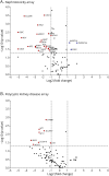
Results: Patients with SMA had alterations in serum creatinine, cystatin C, sodium, glucose, and calcium concentrations, granular casts and crystals in urine, and nephrocalcinosis and fibrosis. Nephrotoxicity and polycystic kidney disease PCR arrays revealed multiple differentially expressed genes, and immunoblot analysis showed decreased calcium-sensing receptors and calbindin and increased insulin-like growth factor-binding proteins in kidneys from patients with SMA.
Conclusions: These findings demonstrate that patients with SMA type 1, in the absence of disease-modifying therapies, frequently manifest impaired renal function as a primary or secondary consequence of their disease. This study provides new insights into systemic contributions to SMA disease pathogenesis and the need to identify coadjuvant therapies.
Figures


References
-
- Groen EJN, Talbot K, Gillingwater TH. Advances in therapy for spinal muscular atrophy: promises and challenges. Nat Rev Neurol 2018;14:214–224. - PubMed
-
- Faravelli I, Nizzardo M, Comi GP, Corti S. Spinal muscular atrophy-recent therapeutic advances for an old challenge. Nat Rev Neurol 2015;11:351–359. - PubMed
-
- Thomas NH, Dubowitz V. The natural history of type I (severe) spinal muscular atrophy. Neuromuscul Disord 1994;4:497–502. - PubMed
-
- Corey DR. Nusinersen, an antisense oligonucleotide drug for spinal muscular atrophy. Nat Neurosci 2017;20:497–499. - PubMed
-
- Wood MJA, Talbot K, Bowerman M. Spinal muscular atrophy: antisense oligonucleotide therapy opens the door to an integrated therapeutic landscape. Hum Mol Genet 2017;26:R151–R159. - PubMed
Grants and funding
LinkOut - more resources
Full Text Sources
