. 2019 Oct 16;11(41):37381-37396.
doi: 10.1021/acsami.9b07053. Epub 2019 Oct 7.

Triple PLGA/PCL Scaffold Modification Including Silver Impregnation, Collagen Coating, and Electrospinning Significantly Improve Biocompatibility, Antimicrobial, and Osteogenic Properties for Orofacial Tissue Regeneration

Affiliations

Triple PLGA/PCL Scaffold Modification Including Silver Impregnation, Collagen Coating, and Electrospinning Significantly Improve Biocompatibility, Antimicrobial, and Osteogenic Properties for Orofacial Tissue Regeneration

Yunzhu Qian et al. ACS Appl Mater Interfaces. .

Abstract

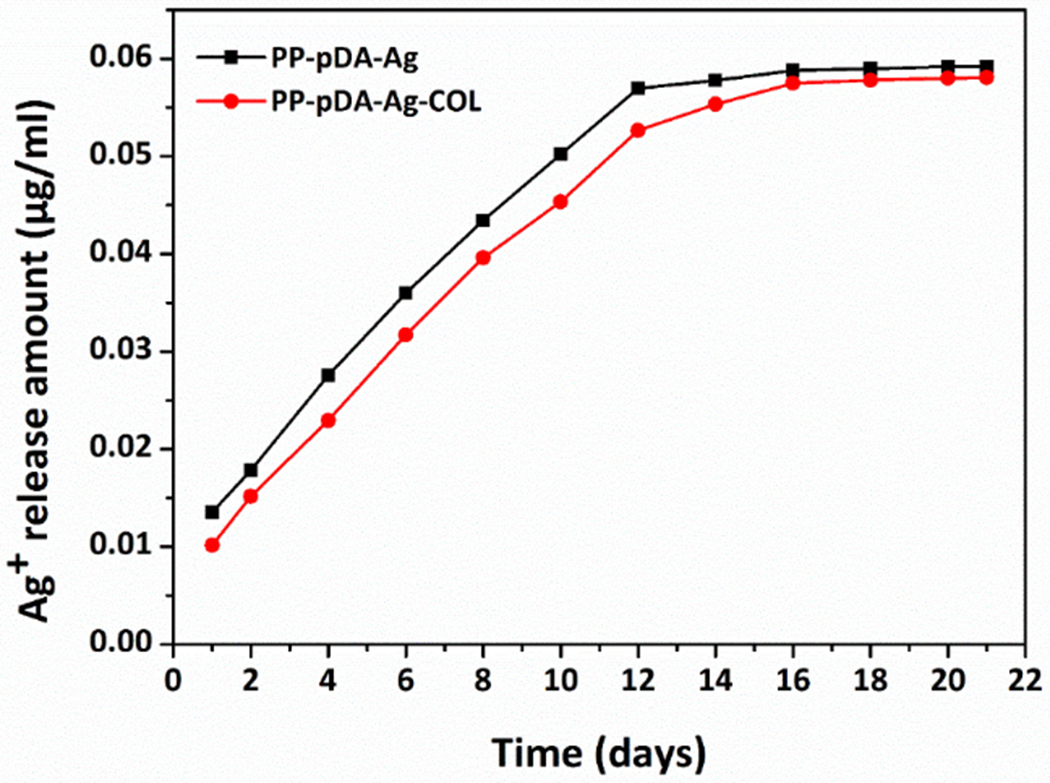
Biodegradable synthetic scaffolds hold great promise for oral and craniofacial guided tissue regeneration and bone regeneration. To overcome the limitations of current scaffold materials in terms of osteogenic and antimicrobial properties, we have developed a novel silver-modified/collagen-coated electrospun poly-lactic-co-glycolic acid/polycaprolactone (PLGA/PCL) scaffold (PP-pDA-Ag-COL) with improved antimicrobial and osteogenic properties. Our novel scaffold was generated by electrospinning a basic PLGA/PCL matrix, followed by silver nanoparticles (AgNPs) impregnation via in situ reduction, polydopamine coating, and then coating by collagen I. The three intermediate materials involved in the fabrication of our scaffolds, namely, PLGA/PCL (PP), PLGA/PCL-polydopamine (PP-pDA), and PLGA/PCL-polydopamine-Ag (PP-pDA-Ag), were used as control scaffolds. Scanning electron micrographs and mechanical testing indicated that the unique three-dimensional structures with randomly oriented nanofibrous electrospun scaffold architectures, the elasticity modulus, and the tensile strength were maintained after modifications. CCK-8 cell proliferation analysis demonstrated that the PP-pDA-Ag-COL scaffold was associated with higher MC3T3 proliferation rates than the three control scaffolds employed. Scanning electron and fluorescence light microscopy illustrated that PP-pDA-Ag-COL scaffolds significantly enhanced MC3T3 cell adhesion compared to the control scaffolds after 12 and 24 h culture, in tandem with the highest β1 integrin expression levels, both at the mRNA level and the protein level. Alkaline phosphatase activity, BMP2, and RUNX2 expression levels of MC3T3 cells cultured on PP-pDA-Ag-COL scaffolds for 7 and 14 days were also significantly higher when compared to controls (P < 0.001). There was a wider antibacterial zone associated in PP-pDA-Ag-COL and PP-pDA-Ag scaffolds versus control scaffolds (P < 0.05), and bacterial fluorescence was reduced on the Ag-modified scaffolds after 24 h inoculation against Staphylococcus aureus and Streptococcus mutans. In a mouse periodontal disease model, the PP-pDA-Ag-COL scaffold enhanced alveolar bone regeneration (31.8%) and was effective for periodontitis treatment. These results demonstrate that our novel PP-pDA-Ag-COL scaffold enhanced biocompatibility and osteogenic and antibacterial properties and has therapeutic potential for alveolar/craniofacial bone regeneration.

Keywords: antimicrobial; biomimetic scaffolds; electrospinning; local delivery; osteogenic; silver nanoparticle.


Conflict of interest statement

The authors declare no competing financial interest.

Figures

Figure 1
Figure 1
Schematic illustration of the preparation procedure of PP-pDA-Ag-COL scaffolds. PLGA/PCL scaffolds were prepared by electrospun technology. Ag nanoparticles were in site reduced by polydopamine, and then coated by collagen I.
Figure 2.
Figure 2.
Structure and composition of the PP-pDA-Ag-COL and control scaffolds. (A, a- D, d) SEM images of the PP (A, a), PP-pDA (B, b), PP-pDA-Ag (C, c) and PP-pDA-Ag-COL (D, d) scaffolds at lower (A, B, C and D) and higher (a, b, c and d) magnification. Note that all four scaffolds exhibited unique 3D architectures with interconnected randomly-oriented nanofibers and mimicked the arrangement of native extracellular matrices. (As-Ds) EDX spectrum of the PP (As), PP-pDA (Bs), PP-pDA-Ag (Cs) and PP-pDA-Ag-COL (Ds) scaffolds, and (E) elemental composition of the scaffold surfaces C-Carbon, O-Oxygen, Ag-Silver.
Figure 3.
Figure 3.
Physicochemical and mechanical properties of the PP-pDA-Ag-COL and control scaffolds. (A). FT-IR spectra of the PP, PP-pDA, PP-pDA-Ag and PP-pDA-Ag-COL scaffolds. (B). Mechanical properties of tensile strength and elastic modulus of the PP, PP-pDA, PP-pDA-Ag and PP-pDA-Ag-COL scaffolds. The elastic modulus (left Y axis) is compared among the four scaffolds by column chart while the tensile strength (right Y axis) is compared by the line chart. (C). Contact angle of the PP, PP-pDA, PP-pDA-Ag and PP-pDA-Ag-COL scaffolds. The images above each column illustrate the actual contact angle when water drops onto the scaffolds. Note the significantly lower contact angle of the PP-pDA-Ag-COL scaffold, indicative of better hydrophilic properties than the three control scaffolds.
Figure 4.
Figure 4.
Cumulative Ag+ release from two silver-modified scaffolds, PP-pDA-Ag and PP-pDA-Ag-COL. Silver ions were accumulatively and steadily released from PP-pDA-Ag and PP-pDA-Ag-COL and measured at 1, 2, 4, 6, 8, 10, 12, 14, 16, 18, 20 and 21 days in PBS buffer. Note that PP-pDA-Ag and PP-pDA-Ag-COL showed similar release curves while the PP-pDA-Ag-COL scaffold had slightly slower release rate.
Figure 5.
Figure 5.
The effect of the PP-pDA-Ag-COL and control scaffolds on cell proliferation. MC3T3 cells were seeded onto the PP, PP-pDA and AgNO3 coated PP-pDA-Ag scaffolds at concentrations of 5×10⁻⁵, 1×10⁻⁴, 5×10⁻⁴ and 1×10⁻³M (A), or the cells were cultured on the PP, PP-pDA, PP-pDA-Ag (10−4M) and PP-pDA-Ag-COL (10−4M) scaffolds (B) for 1, 3, 5 and 7 days. Cell viability and proliferation was determined as OD value and compared with PP using a CCK-8 kit. * p < 0.05, ** p < 0.01, *** p < 0.001.
Figure 6
Figure 6
Fluorescent images of live/dead staining of MC3T3-E1 on scaffolds after culturing for 3 days. Scale bar is 500μm. Live cells were stained in green while dead cells were stained in red.
Figure 7.
Figure 7.
Cell morphology and attachment on the PP-pDA-Ag-COL and control scaffolds. MC3T3 cells were cultured on the PP, PP-pDA, PP-pDA-Ag and PP-pDA-Ag-COL scaffolds for 12h or 24h. (A) FESEM images of the scaffolds with cultured cells for 24h. The arrowheads point to the attached and spread cells on the scaffolds. (B) Fluorescent staining of cytoskeleton after the cells were cultured for 12h and 24h. Note that the MC3T3 cells on the PP-pDA-Ag-COL scaffold had higher cell density and were well spread.
Figure 8.
Figure 8.
Integrins β1, α1, α2, α5, α10, α11 expression in MC3T3 cells cultured on PP, PP-pDA, PP-pDA-Ag and PP-pDA-Ag-COL scaffolds for 12h, 24h and 48h. The expression of target genes was normalized to GAPDH and the relative expression level was calculated by the 2−ΔΔCt method. * p < 0.05, ** p < 0.01, *** p < 0.001, compare with PP.
Figure 9.
Figure 9.
Integrin β1 expression in MC3T3 cells cultured on PP-pDA-Ag-COL and control scaffolds. MC3T3 cells were cultured on the scaffolds for 24 h and then fluorescently labeled for F-actin (red) or for integrin β1 via immunofluorescence (green). Note the increased levels of integrin β1 on the MC3T3 cells on the PP-pDA-Ag-COL scaffold.
Figure 10.
Figure 10.
Effects of the PP-pDA-Ag-COL and control scaffolds on osteogenic differentiation of MC3T3 cells. Cells were cultured on the scaffolds for 7 (A, C, D) or 14 (B, E, F) days. (A, B) ALP activity was determined using an alkaline phosphatase assay kit and normalized to the total protein content. The protein expression of BMP2 and RUNX2 for 7 (C, D) and 14 (E, F) days was determined by Western blotting. The expression levels were calculated by densitometry analysis relative to GAPDH. * p < 0.05, ** p < 0.01, *** p < 0.001, compare with PP.
Figure 11.
Figure 11.
Antibacterial properties of the PP-pDA, PP-pDA-Ag and PP-pDA-Ag-COL scaffolds. (A, B) Agar diffusion tests. Bacteria S. aureus (A) and S. mutans (B) were inoculated in LB agar (A) or Brian Heart Infusion agar (B). The PP (#1), PP-pDA (#2), PP-pDA-Ag (#3) and PP-pDA-Ag-COL (#4) scaffolds were punched into 6 mm disks and placed on the surface of the agars. The plates were incubated for 24 h. The circles point to the diameter of the inhibition zone (DIZ). (C-F) Bacterial attachment on the scaffolds. The PP, PP-pDA, PP-pDA-Ag and PP-pDA-Ag-COL scaffolds were incubated with S. aureu and S. mutans suspension at 37 °C for 24h and then dehydrated. Surface topography and antibacterial ability of the scaffolds was observed by FESEM (C, D). Scar bar= 2 μm. Bacterial fluorescence (E, F) was captured by CLSM after the bacteria were stained with DAPI. Scar bar= 20 μm.
Figure 12.
Figure 12.
Subcutaneous implantation of PP-pDA-Ag-COL and three control scaffolds. (A-D) H&E staining. Note that much more cells penetrated into PP-pDA-Ag-COL (D) scaffolds than PP (A), PP-pDA (B), PP-pDA-Ag (C) scaffolds, indicating that PP-pDA-Ag-COL had better biocompatibility and biomimetic capacity to accommodate cell recruitment. (E-H) Mallory’s Trichrome staining. Collagen fibers, reticulum and mucous shades were stained in blue, elastic fibers in pink, yellow or unstained, muscle, alpha and beta cells in blue and nuclei in red.
Figure 13.
Figure 13.
Effect of our PP-pDA-Ag-COL scaffold on periodontal status following implantation in a periodontitis mouse model. (A) μCT 3D reconstruction of the left maxillary mouse molars after 6-week implantation of PP, PP-pDA-Ag and PP-pDA-Ag-COL scaffolds subjected to GTR treatment. The blank control is the periodontitis mouse model. (B) H&E staining of the regenerated periodontal tissues after 6-week implantation of PP, PP-pDA-Ag and PP-pDA-Ag-COL scaffolds subjected to GTR treatment. The periodontitis mouse model is the control. (C) Alveolar bone loss of the maxillary mouse molars in periodontitis control, PP, PP-pDA-Ag and PP-pDA-Ag-COL. (D) The bone volume in periodontitis control, PP, PP-pDA-Ag and PP-pDA-Ag-COL. (E) Bone mineral density of the alveolar bone in periodontitis control, PP, PP-pDA-Ag and PP-pDA-Ag-COL. * p < 0.05, *** p < 0.001 multiple compare. There was significantly less bone loss in the PP-pDA-Ag-COL scaffold group, suggesting that the local release of AgNPs delivered by the novel scaffolds PP-pDA-Ag-COL via GTR treatments contributed to the alleviation of periodontal inflammation and facilitated bone regeneration in a periodontitis mouse model.

References

    1. Bottino MC; Thomas V; Schmidt G; Vohra YK; Chu TMG; Kowolik MJ; Janowski GM Recent advances in the development of GTR/GBR membranes for periodontal regeneration-A materials perspective. Dent. Mater. 2012, 28 (7), 703–721, DOI: 10.1016/j.dental.2012.04.022. - DOI - PubMed
    1. Soldatos NK; Stylianou P; Koidou VP; Angelov N; Yukna R; Romanos GE Limitations and options using resorbable versus nonresorbable membranes for successful guided bone regeneration. Quintessence Int. 2017, 48 (2), 131–147. - PubMed
    1. Huang GY; Li F; Zhao X; Ma YF; Li YH; Lin M; Jin GR; Lu TJ; Genin GM; Xu F Functional and Biomimetic Materials for Engineering of the Three-Dimensional Cell Microenvironment. Chem. Rev. 2017, 117 (20), 12764–12850, DOI: 10.1021/acs.chemrev.7b00094. - DOI - PMC - PubMed
    1. Qian YZ; Zhou XF; Sun H; Yang JX; Chen Y; Li C; Wang HJ; Xing T; Zhang FM; Gu N Biomimetic Domain-Active Electrospun Scaffolds Facilitating Bone Regeneration Synergistically with Antibacterial Efficacy for Bone Defects. ACS Appl. Mater. Interfaces 2018, 10 (4), 3248–3259, DOI: 10.1021/acsami.7b14524. - DOI - PubMed
    1. Wang JL; Wang LN; Zhou ZY; Lai HJ; Xu P; Liao L; Wei JC Biodegradable Polymer Membranes Applied in Guided Bone/Tissue Regeneration: A Review. Polymers 2016, 8 (4), 20, DOI: 10.3390/polym8040115. - DOI - PMC - PubMed

MeSH terms