

doi: 10.1364/OL.44.004451.
Solid immersion meniscus lens (SIMlens) for open-top light-sheet microscopy
- PMID: 31517904
- PMCID: PMC7331451
- DOI: 10.1364/OL.44.004451
Item in Clipboard
Solid immersion meniscus lens (SIMlens) for open-top light-sheet microscopy
Opt Lett.
.
Abstract
Open-top light-sheet (OTLS) microscopy has been developed for rapid volumetric imaging of large pathology specimens. A challenge with OTLS microscopy is the transmission of oblique illumination and detection beams through a horizontal sample plate without introducing aberrations. Previous solutions prevented vertical sample movement, constrained the refractive index of the sample, and/or hindered multi-resolution imaging. Here we describe a solid immersion meniscus lens, a wavefront-matching element that suppresses aberrations when illumination and detection beam transition between air and various high-index immersion media, thereby enabling multi-resolution OTLS microscopy of specimens cleared with diverse protocols.
Figures

(a) An OTLS microscope rapidly images a sample in a plane-by-plane fashion. (b) Our first demonstration of OTLS microscopy utilized a SIL, positioned beneath a glass plate, in which the curved outer surface of the SIL was perpendicular to the optical rays (i.e., wavefront-matching), as they transitioned between air and glass, thereby preventing refractive aberrations as would occur at a flat angled interface.

(a) A SIMlens in front of an air objective. The radii of curvature of both SIMlens surfaces are matched to the objective’s focal point, such that rays emanating from the focal point do not refract as they cross both SIMlens interfaces. Since ray angles (θ) are pre served, the NA and magnification increase by a factor of n, the refractive index of the immersion medium. (b) In OTLS microscopy, a SIMlens used with an air-based objective suppresses off-axis and spherical aberrations. This architecture accommodates a variety of immersion media and enables the rapid exchange of objectives for multi-resolution imaging.

(a) Optical setup to demonstrate compatibility of a SIMlens with various immersion media. A resolution target is back-illuminated in an air or liquid medium (n = 1.00, 1.33, or 1.56). The detection arm consists of a SIMlens (secured to the chamber with a gasket and retaining ring), air objective (10× or 20×), tube lens, and camera. (b) Setup to demonstrate resolution uniformity when using a SIMlens on the detection arm of a light-sheet microscope. A light sheet illuminates a sample immersed in a liquid bath. The fluorescence signal is imaged onto a camera with the same setup used in (a).

(a) Images of a resolution target within media of different refractive indices. The images were obtained with a SIMlens in conjunction with: (a) a 10× objective or (b) a 20× air objective. Note that magnification increases with n, but the images are cropped to show the same FOV. (c) As expected, the resolution (Rayleigh criterion) measured at the center of the FOV improves as n increases (resolution ∝ 1∕n) and is measured to be within 10% and 30% of the diffraction limit when using the 10× and 20× objective, respectively.

Sub-diffraction beads were imaged to assess the resolution across the FOV. (a) Measured lateral resolution (FWHM of main lobe) across the FOV is plotted for a 10× objective. The use of a SIMlens with an immersion medium at n = 1.56 results in an effective NA (NAeff) of 0.33. The measurement uncertainty (standard deviation) is indicated by the shaded region. Also shown are theoretical results based on Zemax simulations. Representative bead images are shown as acquired from the left, center, and right edges of the FOV. (b) Similar results are shown for a 20× air objective (NAeff = 0.62). The scale bars are 5 μm.
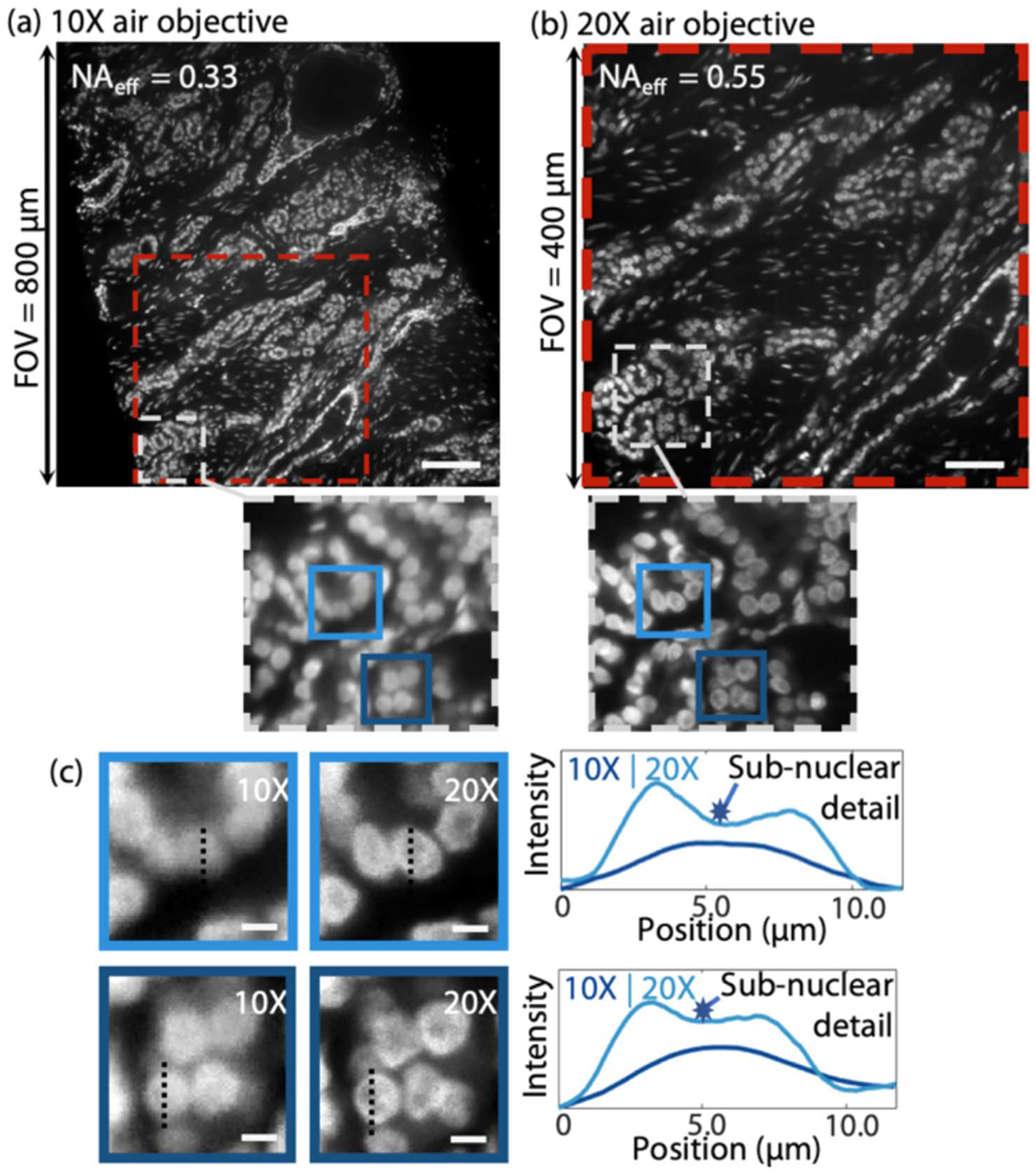
(a) Low-magnification view of human prostate tissue cleared and index matched at n = 1.56, fluorescently stained with TO-PRO3, and imaged with a SIMlens and 10× air objective (NAair = 0.21, NAeff = 0.33). The scale bar is 100 μm. (b) Same region of the tissue imaged at higher magnification, with a 20× air objective (NAair = 0.35, NAeff = 0.55) and a SIMlens. The scale bar is 50 μm. (c) Line profiles across individual nuclei demonstrate the resolution improvement achieved with a 20× objective compared to a 10× objective. Nucleoli are apparent in images obtained with the 20× objective (dark central regions within the nuclei), but are not resolved with the 10× objective. The scale bars are 5 μm.
References
Grants and funding
LinkOut - more resources
Full Text Sources
Other Literature Sources
