. 2019 Nov 1;79(21):5626-5639.
doi: 10.1158/0008-5472.CAN-19-0800. Epub 2019 Sep 13.

Neutrophil Extracellular Traps Drive Mitochondrial Homeostasis in Tumors to Augment Growth

Affiliations

Neutrophil Extracellular Traps Drive Mitochondrial Homeostasis in Tumors to Augment Growth

Hamza O Yazdani et al. Cancer Res. .

Abstract

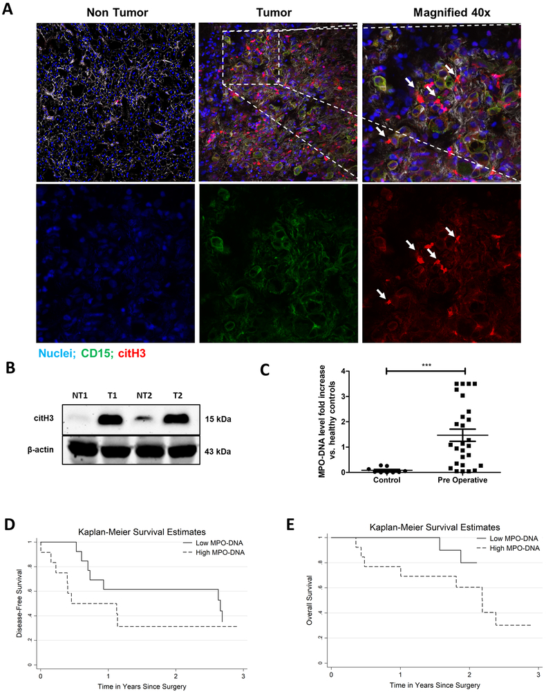
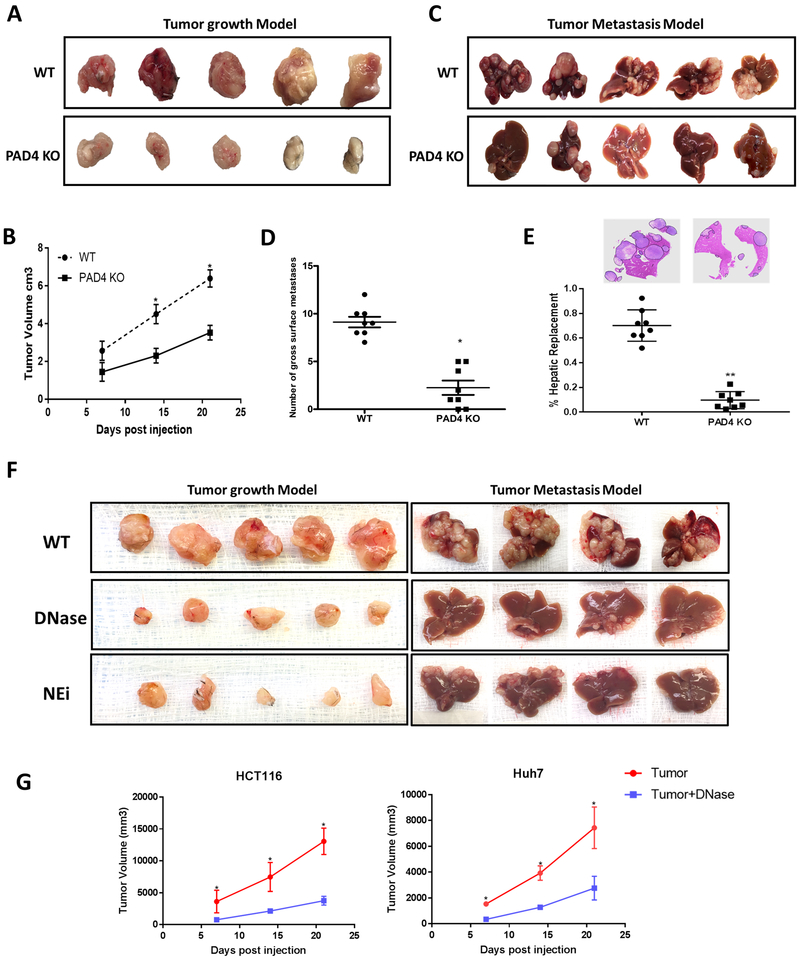
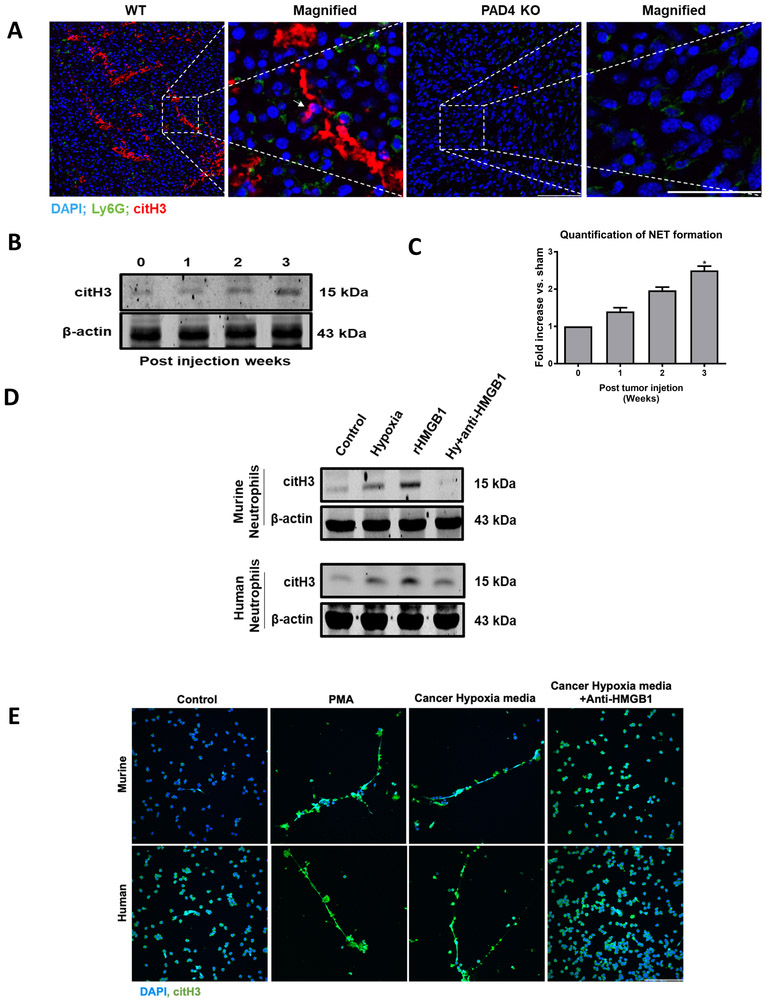
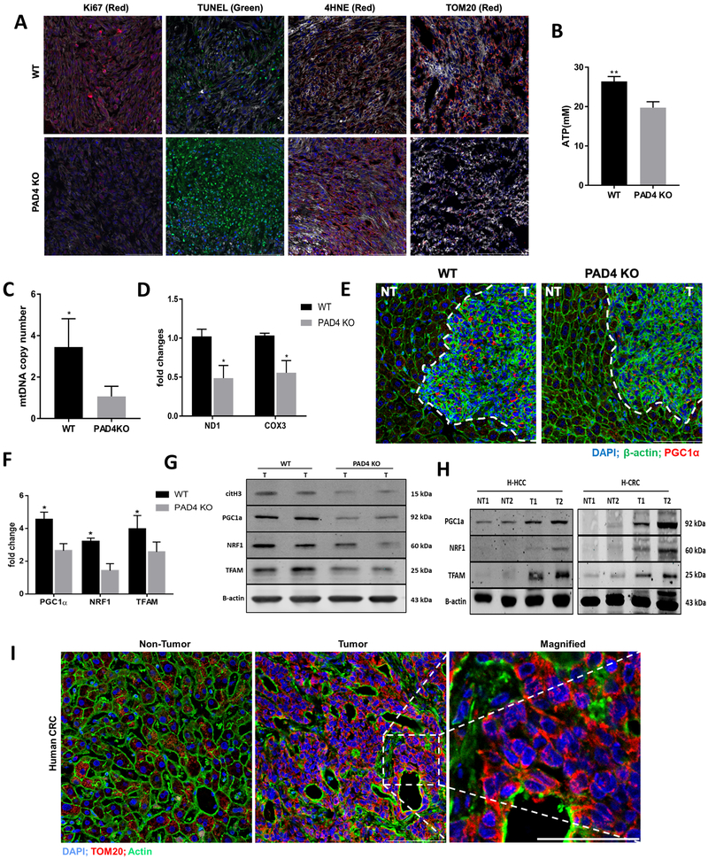
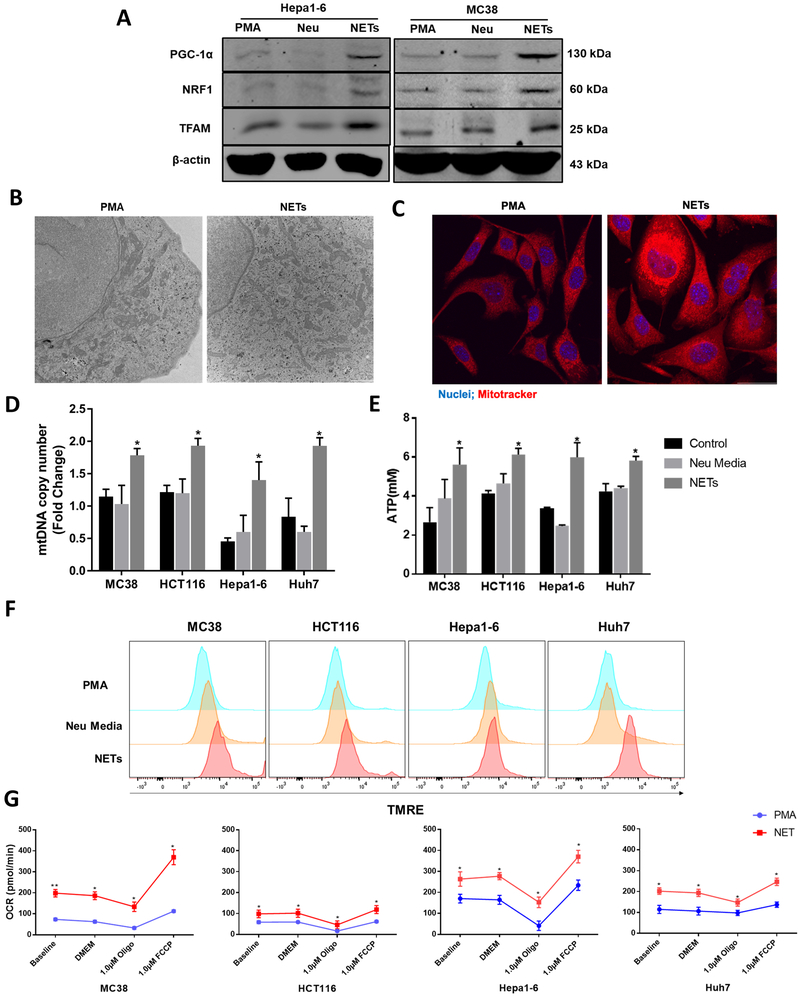
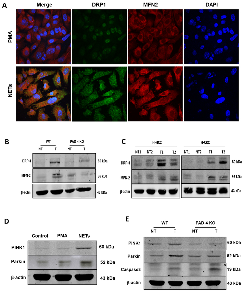
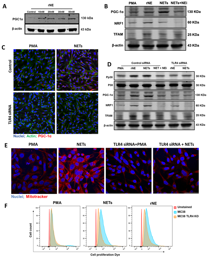
Neutrophil infiltration and neutrophil extracellular traps (NET) in solid cancers are associated with poorer prognosis, but the mechanisms are incompletely understood. We hypothesized that NETs enhance mitochondrial function in tumor cells, providing extra energy for accelerated growth. Metastatic colorectal cancer tissue showed increased intratumoral NETs and supranormal preoperative serum MPO-DNA, a NET marker. Higher MPO-DNA correlated with shorter survival. In mice, subcutaneous tumor implants and hepatic metastases grew slowly in PAD4-KO mice, genetically incapable of NETosis. In parallel experiments, human cancer cell lines grew slower in nu/nu mice treated with DNAse, which disassembles NETs. PAD4-KO tumors manifested decreased proliferation, increased apoptosis, and increased evidence of oxidative stress. PAD4-KO tumors had decreased mitochondrial density, mitochondrial DNA, a lesser degree of ATP production, along with significantly decreased mitochondrial biogenesis proteins PGC1α, TFAM, and NRF-1. In vitro, cancer cells treated with NETs upregulated mitochondrial biogenesis-associated genes, increased mitochondrial density, increased ATP production, enhanced the percentage of cancer cells with reduced mitochondrial membrane potential, and increased the oxygen consumption rate. Furthermore, NETs increased cancer cells' expression of fission and fusion-associated proteins, DRP-1 and MFN-2, and mitophagy-linked proteins, PINK1 and Parkin. All of which were decreased in PAD4-KO tumors. Mechanistically, neutrophil elastase released from NETs activated TLR4 on cancer cells, leading to PGC1α upregulation, increased mitochondrial biogenesis, and accelerated growth. Taken together, NETs can directly alter the metabolic programming of cancer cells to increase tumor growth. NETs represent a promising therapeutic target to halt cancer progression. SIGNIFICANCE: Neutrophils through the release of NETs facilitate the growth of stressed cancer cells by altering their bioenergetics, the inhibition of which induces cell death.


Conflict of interest statement

Conflict of Interest: No potential conflicts of interest were disclosed by the authors

Figures

Figure 1.
Figure 1.. NET formation correlates with cancer specific outcomes in patients with metastatic colorectal cancer.
A, Representative immunofluorescence images by confocal microscopy of human colorectal liver metastases (CRLM) tissue sections showing increased neutrophil infiltration and neutrophil extracellular trap (NET) formation in tumor at 20× magnification compared to non-tumor tissue of the same patient. White arrows showing neutrophils releasing NETs in merge and single staining magnified images at 40×, scale bar 50 µm. B, Protein citH3 levels were increased as evident by western blot image between the tumor (T) and non-tumor (NT) tissue of human CRLM. The blot shown is representative of three independent experiments with similar results. C, Pre-operative MPO-DNA levels detected by Elisa kit are significantly higher in patients with CRLM (n=27) compared to healthy volunteers (n=10, ***p<0.0002). D and E, Kaplan-Meier disease-free and overall survival curves were based on high versus low MPO-DNA levels post-operatively for three years (log-rank test p<0.001 for both D and E).
Figure 2.
Figure 2.. Tumor growth is reduced in neutrophil extracellular traps (NETs) depleted mice.
Subcutaneous tumors using MC38 cells (1×10⁶) injected subcutaneously (A) or through the spleen (C) for the metastasis model showing smaller tumors harvested 3 weeks post-inoculation in PAD4 KO compared to WT mice (n=5/group). B and D, Graphs showing significantly decreased tumor volume and surface liver nodules, respectively, in PAD4 KO mice compared to WT control *P <0.05. E, Hematoxylin and Eosin (H&E) staining of liver sections exhibit decreased tumor burden in PAD4 KO mice (n=5) compared to WT **P <0.01. F, Similarly, mice treated daily with DNAse (50ug) or Neutrophil Elastase inhibitor (NEi) (2.0 mg/kg) in both subcutaneous (n=5) and metastatic model (n=4) showed decrease tumor growth compared to controls. G, Graph representing tumor growth curve in DNAse (50ug) treated Nu/Nu athymic mice inoculated with HCT116 and Huh7 cell lines (1×10⁶) (n=5/group). Data presented as mean SEM. *P <0.05.
Figure 3.
Figure 3.. Solid tumors recruit neutrophils and induce NET formation in their microenvironment.
A, Sectional staining of harvested tumor tissue showing neutrophils and neutrophil extracellular traps (NETs) infiltration three weeks after the cancer injection in WT and PAD4 KO mice (magnification 40×, magnified image 60×, scale bar, 50 µm). White arrow showing neutrophils releasing NETs. Nucleus (blue), Ly6G (green) and citH3 (red). B and C, CitH3 protein expression by western blot and Elisa quantification of serum MPO-DNA level (n=3/group) in growing tumors was assessed at indicated time. Invitro, neutrophils isolated from healthy human volunteer (blood) or mouse (bone marrow) when cocultured with 24h hypoxic cancer media (HCT116 and MC38) and rHMGB1 (1µg) for 4h resulted in an increase NET formation as evident by western blot (D) and confocal microscopy (E) at 40× magnification. Neutrophils treated with PMA (250 nM) serves as a positive control. Anti-HMGB1 antibody added to cancer hypoxic media inhibited NET formation when cocultured with neutrophils. Abbreviations Hy, hypoxia. The blots shown are representatives of three independent experiments with similar results. *P <0.05.
Figure 4.
Figure 4.. NETs in the tumor microenvironment correlate with evidence of increased mitochondrial biogenesis and more rapid tumor growth.
A, A marked decrease in cell proliferation rate (Ki67) and increased apoptosis (TUNEL) was observed in PAD4 KO tumors. Ki67+ (median 25 [3-44] Ki67+ cells/106 µm2 in PAD4KO tumor tissue versus 120 [62-159] Ki67+ cells/106 µm2 in WT and TUNEL (median 31 [5-49.3] TUNEL+ cells/103 µm2 in PAD4KO tumor tissue versus 17 [9-35] TUNEL+ cells/103 µm2 in WT, P <0.05). Increase in the oxidative stress observed by positive 4-HNE staining in PAD4 KO tissue (median 0.193 [0.067-0.438] in PAD4 KO versus 0.053 [0.005-0.079] in WT control, P<0.05. PAD4 KO tumors showed impaired mitochondrial density compared with WT tumors when stained for TOM20. The median normalized TOM20+ area in PADKO tumor tissue was 0.087 (range 0.0065-0.197) versus 0.438 (range 0.292-0.504) in control tumors. Nuclei (blue), actin (white), Ki67/4HNE/TOM20 (red), TUNEL (green). Scale bars, 100 µm, n=5 mice/group. Values were based on analysis of tumors from five mice for each group. There was a significant decrease in mtDNA copy numbers measured by RT-PCR (B) and lesser degree of ATP levels (C) in tumors lacking NETs. **P < 0.01, *P < 0.05. D, COX3 and ND1 expression was analyzed by western blot showing significantly low in protein density, *P < 0.05. E, Three weeks after splenic injection the liver tumor area of WT mouse showing upregulated PGC-1α expression as evident by immunofluorescence comparing PAD4 KO. No differences in the non-tumor section of both mice were observed. Magnification 40×, scale bars 50 µm. F and G, Genes associated with mitochondrial biogenesis were assessed by both Real-time PCR and western blot in the tumor tissue of PAD4 KO and WT mice, *P < 0.05. H, Protein expression of citH3, PGC-1α, NRF1, and TFAM in human hepatocellular (H-HCC) and colorectal (H-CRC) tumor tissue (T) comparing its non-tumor (NT) counterpart. The blots shown are representatives of three independent experiments with similar results I, The human colorectal liver metastasis (CRLM) tissue showing an increased mitochondrial density in the tumor section compared to non-tumor area as observed by immunofluorescence staining of TOM20 staining (magnification 40×, magnified 60×, scale bars 50 µm). Abbreviations; TUNEL, terminal deoxynucleotidyl transferase–mediated deoxyuridine triphosphate nick-end labeling.
Figure 5.
Figure 5.. NETs directly alter the metabolism of cancer cells by enhancing mitochondrial function and biogenesis in vitro.
Neutrophils isolated from mice were stimulated with PMA for 4 hours to form NETs. Extracted NETs were cocultured with cancer cells (MC38 and Hepa-1-6) overnight. A, Representative images of proteins expressing PGC-1α, NRF1 and TFAM by western blot in indicated groups. The blots shown are representatives of three independent experiments with similar results. B and C, Electron microscopy and confocal microscopy of NETs treated MC38 cells showing significant increase in the number of mitochondria within the cells (magnification 10×, scale bar 50 µm) P<0.05. Mitotracker (red), nuclei (blue). The Real-Time PCR of mtDNA copy number (D) and the production of intracellular ATP levels (E) was observed in both human and mouse cancer cells between groups treated with PMA, neutrophil media and NETs, *P < 0.05. F, As witnessed by the staining of Tetramethylrhodamine ethyl ester (TMRE) flowcytometry analysis showing increased mitochondrial membrane potential following NET treatment in cancer cells. G, The mitochondrial respiratory capacity of cancer cells was elevated in the NET treated group when cell oxygen consumption rate (OCR) was tested, *P < 0.05, **P < 0.01. PMA (250nM) group was added to serve as a control group in each experiment.
Figure 6.
Figure 6.. NETs preserve mitochondrial homeostasis and dynamics: fission, fusion and mitophagy.
A, After PMA stimulation for 4h, neutrophil extracellular traps (NETs) were cocultured with MC38 cancer cell line and confocal microscopy was utilized to analyze proteins regulating mitochondrial fission and fusion. NET treatment upregulated the expression of DRP1 and MFN2 proteins compared to cells treated with PMA as a control, nuclei (blue), DRP1 (green), and MFN2 (red). magnification 40× scale bar 50 µm. B, 3 weeks after the splenic injection the liver tumor tissue of PAD4 KO mice showing downregulated expression of DRP1 and MFN2 compared to its non-tumor background. Similarly, human hepatocellular (H-HCC) and human colorectal (H-CRC) tumor (T) sections showing upregulation in these proteins comparing non-tumor (NT) counterparts. D, Representative western blot analysis image showing increased induction of mitophagy as evident by the upregulation in the expression of protein PINK1 and Parkin in MC38 cancer cells when treated with NETs. E, Parallel results can be seen in the tumor tissues of the WT mice 3 weeks after injection as analyzed by western blot. The blots shown are representatives of three independent experiments with similar results.
Figure 7.
Figure 7.. Neutrophil Elastase released by NETs induces mitochondrial biogenesis through TLR4-p38-PGC1-α pathway in MC38 cells.
A, Western blot analysis showing expression of protein PGC-1α in cancer cells treated endogenously with recombinant neutrophil elastase (rNE) at indicated concentration. B, Addition of NETs or rNE (30nM) in the culturing cancer cells increased the expression of PGC-1α, NRF1 and TFAM when determined. However, the addition of neutrophil elastase inhibitor (NEi) together with NETs inhibited this effect. C, Immunofluorescence staining of TLR4 transfected cells cocultured with NETs showing decreased PGC-1a expression with the staining of nuclei (blue) PGC-1α (red) and actin (green). Magnification 20×, scale bar 50 µm. D, Western blot analysis showing downregulated expression of phospho-p38, PGC-1α, NRF1, and TFAM when treated with rNE (30nM) and NETs in TLR4 transfected cells. The blots shown are representatives of three independent experiments with similar results. E, Intracellular mitochondria staining was observed with Mitotracker (red) staining between the groups Magnification 40×, scale bar 50 µm. F, Flowcytometry analysis showing increased cancer cell proliferation when stained with proliferation dye 48h after treating with isolated NETs or rNE. PMA (250nM) group was added to serve as a control group in each experiment.

References

    1. Gajewski TF, Schreiber H, Fu YX. Innate and adaptive immune cells in the tumor microenvironment. Nat. Immunol. 2013. - PMC - PubMed
    1. Kahlert C, Pecqueux M, Halama N, Dienemann H, Muley T, Pfannschmidt J, et al. Tumour-site-dependent expression profile of angiogenic factors in tumour-associated stroma of primary colorectal cancer and metastases. Br J Cancer. 2014; - PMC - PubMed
    1. Reid MD, Basturk O, Thirabanjasak D, Hruban RH, Klimstra DS, Bagci P, et al. Tumor-infiltrating neutrophils in pancreatic neoplasia. Mod Pathol. 2011; - PMC - PubMed
    1. Li YW, Qiu SJ, Fan J, Zhou J, Gao Q, Xiao YS, et al. Intratumoral neutrophils: A poor prognostic factor for hepatocellular carcinoma following resection. J Hepatol. 2011; - PubMed
    1. J TO, S H, M HJ, D F, H M, S P, et al. Intratumoral neutrophils, plasmacytoid dendritic cells, and pSTAT3 in AJCC stage I/II melanoma prognosis. J. Clin. Oncol. 2011. - PubMed

Publication types

MeSH terms