. 2019 Nov 1;294(44):16228-16240.
doi: 10.1074/jbc.RA119.009543. Epub 2019 Sep 13.

Human cardiac myosin-binding protein C restricts actin structural dynamics in a cooperative and phosphorylation-sensitive manner

Affiliations

Human cardiac myosin-binding protein C restricts actin structural dynamics in a cooperative and phosphorylation-sensitive manner

Thomas A Bunch et al. J Biol Chem. .

Abstract

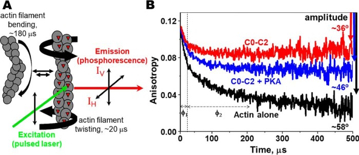
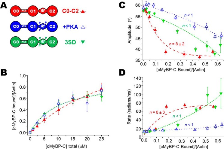
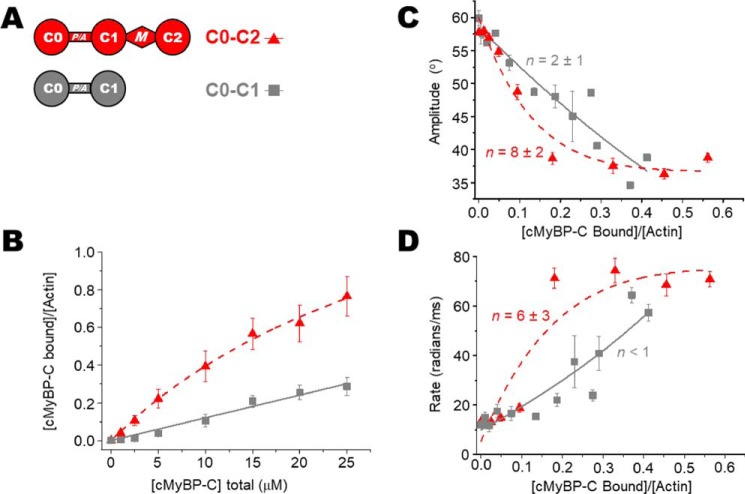
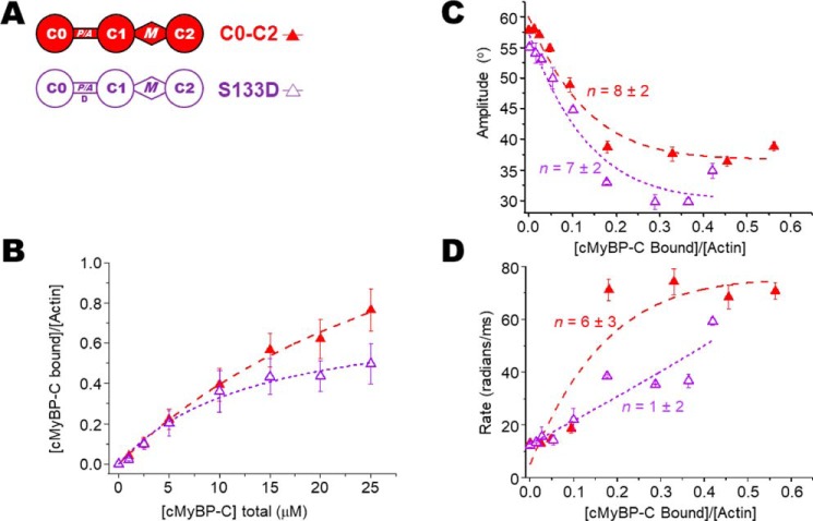
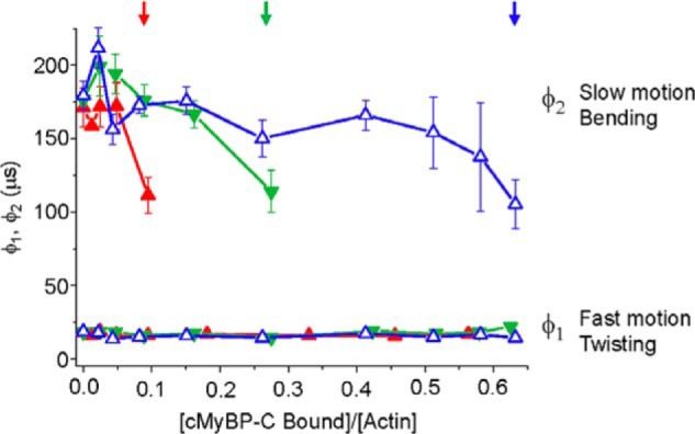
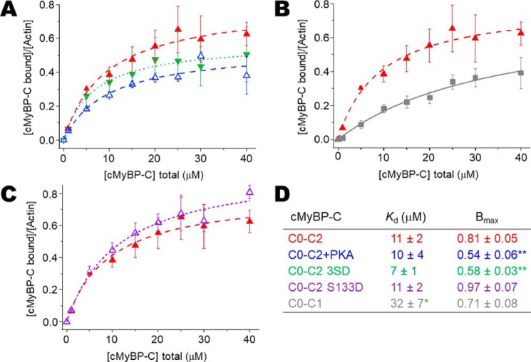
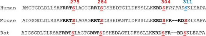
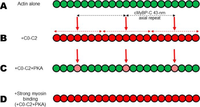
Cardiac myosin-binding protein C (cMyBP-C) is a thick filament-associated protein that influences actin-myosin interactions. cMyBP-C alters myofilament structure and contractile properties in a protein kinase A (PKA) phosphorylation-dependent manner. To determine the effects of cMyBP-C and its phosphorylation on the microsecond rotational dynamics of actin filaments, we attached a phosphorescent probe to F-actin at Cys-374 and performed transient phosphorescence anisotropy (TPA) experiments. Binding of cMyBP-C N-terminal domains (C0-C2) to labeled F-actin reduced rotational flexibility by 20-25°, indicated by increased final anisotropy of the TPA decay. The effects of C0-C2 on actin TPA were highly cooperative (n = ∼8), suggesting that the cMyBP-C N terminus impacts the rotational dynamics of actin spanning seven monomers (i.e. the length of tropomyosin). PKA-mediated phosphorylation of C0-C2 eliminated the cooperative effects on actin flexibility and modestly increased actin rotational rates. Effects of Ser to Asp phosphomimetic substitutions in the M-domain of C0-C2 on actin dynamics only partially recapitulated the phosphorylation effects. C0-C1 (lacking M-domain/C2) similarly exhibited reduced cooperativity, but not as reduced as by phosphorylated C0-C2. These results suggest an important regulatory role of the M-domain in cMyBP-C effects on actin structural dynamics. In contrast, phosphomimetic substitution of the glycogen synthase kinase (GSK3β) site in the Pro/Ala-rich linker of C0-C2 did not significantly affect the TPA results. We conclude that cMyBP-C binding and PKA-mediated phosphorylation can modulate actin dynamics. We propose that these N-terminal cMyBP-C-induced changes in actin dynamics help explain the functional effects of cMyBP-C phosphorylation on actin-myosin interactions.

Keywords: actin; cardiac muscle; cardiac myosin–binding protein C (cMyBP-C); contractile protein; motor protein; myofilament; phosphorylation; protein kinase A (PKA); spectroscopy; structural dynamics.


Conflict of interest statement

The authors declare that they have no conflicts of interest with the contents of this article

Figures

Figure 1.
Figure 1.
Transient phosphorescence anisotropy (TPA) of labeled actin filaments. A, actin filament rotational/twisting (and bending) motions in the microsecond time range are detected by TPA. B, effect of 4 μm C0–C2 (red) or PKA-phosphorylated C0–C2 (blue) on the TPA decay of 1 μm ErIA-actin (black). The vertical dotted line separates the two exponentials of fits for correlation times, φ1 (fast mode, twisting motion, ∼20 μs) and φ2 (slow mode, bending motion, ∼180 μs). Total change in anisotropy (amplitude) is indicated by the downward arrows. The calculated angular amplitude of actin (from Equation 3) is shown. Further details are provided in “Experimental procedures.”
Figure 2.
Figure 2.
Effects of PKA phosphorylation and phosphomimetic substitutions in M-domain of C0–C2 on the TPA of actin. A, domain organization of N-terminal cMyBP-C constructs C0–C2, C0–C2+PKA, and C0–C2 3SD. Domains listed from N-terminal C0 to C2, including the Pro/Ala-rich linker (P/A) and M-domain containing three phosphorylation sites (the P in +PKA) and replacement of the phosphorylated serines with aspartic acid residues (the D in 3SD). B, binding, from cosedimentation experiments, of C0–C2 constructs to 1 μm phalloidin-stablized ErIA-actin. C, angular amplitude (°) was calculated from final anisotropy of actin (Fig. 1B and Equation 3) as a function of v, the fraction bound to N-terminal cMyBP-C. Curves show fits of a cooperativity model to determine n2 = 4, 2, and 11 for C0–C2, C0–C2+PKA, and C0–C2 3SD, respectively. For reference, for linear fits χ2 = 25, 1, and 5, respectively.). D, rates of rotational motions (radians/ms) as a function of v, the fraction bound, and fit to a cooperativity model to determine n. Error bars denote ± S.E.
Figure 3.
Figure 3.
Effects of C0–C1 (lacking M-domain) on the TPA of actin. A, domain organization of N-terminal cMyBP-C constructs C0–C2 and C0–C1. B, binding, from cosedimentation experiments, of C0–C2/C0–C1 constructs to 1 μm phalloidin-stabilized ErIA-actin. C, angular amplitude (°) of actin as a function of v, the fraction bound to N-terminal cMyBP-C. Curves show fits of a cooperativity model to determine n2 = 4 and 6 for C0–C2 and C0-C1, respectively. For reference, for linear fits χ2 = 25 and 6, respectively.). D, rates of rotational motions (radians/ms) as a function of v, the fraction bound and fit to a cooperativity model to determine n. Error bars denote ± S.E.
Figure 4.
Figure 4.
Effects of S133D mutation to mimic P/A linker phosphorylation on the TPA of actin. A, domain organization of N-terminal cMyBP-C constructs C0–C2 and C0–C2 S133D. The Asp for Ser substitution (D) is at the putative GSK3β phosphorylation site in the P/A linker. B, binding of C0–C2 constructs to 1 μm phalloidin-stablized ErIA-actin. C, angular amplitude (°) of actin as a function of v, the fraction bound to N-terminal cMyBP-C. Curves show fits of a cooperativity model to determine n2 = 4 and 9 for C0–C2 and C0–C2 S133D, respectively. For reference, for linear fits χ2 = 25 and 27, respectively.). D, rates of rotational motions (radians/ms) as a function of v, the fraction bound, and fit to a cooperativity model to determine n. Error bars denote ± S.E.
Figure 5.
Figure 5.
Analysis of μs correlation times. The effects of increasing fractions (v) of bound C0–C2 (unphosphorylated (red), 3SD (green), and PKA-phosphorylated (blue)) on the individual correlation times for φ1, the fast mode that captures twisting motions, and φ2, the slow mode that captures bending motions. Note that correlation time is inversely proportional to rate (Equation 4). Arrows indicate the highest binding ratio v of (cMyBP-C bound)/(actin) at which the TPA decays fit best to two exponentials. Higher binding ratios fit best to one exponential—the fast, twisting, motion. Error bars denote ± S.E.
Figure 6.
Figure 6.
Cosedimentation binding data for N-terminal cMyBP-C with unlabeled actin at 5 μm. Although binding data used for TPA analysis were done using phalloidin-stabilized ErIA-labeled F-actin at 1 μm, here nonstabilized and unlabeled F-actin at 5 μm (final concentration) is used to demonstrate that binding of N-terminal cMyBP-C (0–40 μm) with actin under these conditions is consistent with earlier results by others (9, 22, 23). A–C, comparison of C0–C2 (in red in each panel) with modifications and mutations that were used in Figs. 2, 3, and 4, respectively. D, apparent dissociation constants (Kd) and molar binding ratios (Bmax) for binding of recombinant N-terminal cMyBP-C to unlabeled F-actin. Data represent mean ± S.E. Significant difference in Kd or Bmax relative to human C0–C2 (*, p < 0.01; **, p < 0.005). Error bars denote ± S.E.
Figure 7.
Figure 7.
Species-specific cMyBP-C PKA sites. Alignment of human, mouse, and rat sequences within the M-domain that contain PKA recognition sites “R-R-X-S.” Four sites are found in mouse and rat but in human the final target sequence is disrupted by insertion of T-P between the R-R upstream of Ser-311.
Figure 8.
Figure 8.
Actin filament rotational dynamics cooperatively affected by N-terminal cMyBP-C. Actin filaments are illustrated as a line of connected circles (actin monomers). Actin monomers are depicted as having large (58°) angular amplitudes (green) or restricted (36°) angular amplitudes (red). We speculate that these states are representative of weak and strong binding (to myosin) states of actin in cardiac muscle. A, actin alone (all green) exhibits weak-binding dynamics. B, vertical red arrows indicate the approximate position of N-terminal cMyBP-C binding on actin, given the 43-nm axial periodicity of cMyBP-C (horizontal black dashed arrows) in the C-zone of the cardiac muscle sarcomere. Given this spacing, our TPA results are consistent with the idea that adjacent unphosphorylated cMyBP-Cs could propagate structural effects of restricting dynamics all along actin residing in the C-zone. C, PKA treatment limits the spread of the structural effects to only its bound actin monomer. Angular amplitude is only 46° (stippled red in panel C) with phosphorylated C0–C2. D, during β-adrenergic stimulation in cardiac muscle, the increase in myosin interactions with actin would be predicted to dominate the effects on actin structural dynamics, of weak- to strong-binding states, over that of phosphorylated cMyBP-C.

References

    1. Thomas D. D., Muretta J. M., Colson B. A., Mello R. N., and Kast D. (2012) Spectroscopic probes of muscle proteins. in Comprehensive Biophysics (Egelman E. H., ed), Vol. 4, pp. 226–250, Elsevier, Amsterdam: 10.1016/B978-0-12-374920-8.00415-X - DOI
    1. Hopkins S. C., Sabido-David C., van der Heide U. A., Ferguson R. E., Brandmeier B. D., Dale R. E., Kendrick-Jones J., Corrie J. E., Trentham D. R., Irving M., and Goldman Y. E. (2002) Orientation changes of the myosin light chain domain during filament sliding in active and rigor muscle. J. Mol. Biol. 318, 1275–1291 10.1016/S0022-2836(02)00189-4 - DOI - PubMed
    1. Thomas D. D., Seidel J. C., and Gergely J. (1979) Rotational dynamics of spin-labeled F-actin in the sub-millisecond time range. J. Mol. Biol. 132, 257–273 10.1016/0022-2836(79)90259-6 - DOI - PubMed
    1. Prochniewicz E., Walseth T. F., and Thomas D. D. (2004) Structural dynamics of actin during active interaction with myosin: Different effects of weakly and strongly bound myosin heads. Biochemistry 43, 10642–10652 10.1021/bi049914e - DOI - PubMed
    1. Zoghbi M. E., Woodhead J. L., Moss R. L., and Craig R. (2008) Three-dimensional structure of vertebrate cardiac muscle myosin filaments. Proc. Natl. Acad. Sci. U.S.A. 105, 2386–2390 10.1073/pnas.0708912105 - DOI - PMC - PubMed

Publication types