Review
. 2019 Dec;19(12):686-697.
doi: 10.1038/s41568-019-0199-4. Epub 2019 Sep 13.

Cancer biology as revealed by the research autopsy

Affiliations
Review

Cancer biology as revealed by the research autopsy

Christine A Iacobuzio-Donahue et al. Nat Rev Cancer. 2019 Dec.

Abstract

A research autopsy is a post-mortem medical procedure performed on a deceased individual with the primary goal of collecting tissue to support basic and translational research. This approach has increasingly been used to investigate the pathophysiological mechanisms of cancer evolution, metastasis and treatment resistance. In this Review, we discuss the rationale for the use of research autopsies in cancer research and provide an evidence-based discussion of the quality of post-mortem tissues compared with other types of biospecimens. We also discuss the advantages of using post-mortem tissues over other types of biospecimens, including the large amounts of tissue that can be obtained and the extent of multiregion sampling that is achievable, which is not otherwise possible in living patients. We highlight how the research autopsy has supported the identification of the clonal origins and modes of spread among metastases, the extent that selective pressures imposed by treatments cause bottlenecks leading to parallel and convergent tumour evolution, and the creation of rare tissue banks and patient-derived model systems. Finally, we comment on the future of the research autopsy as an integral component of precision medicine strategies.


Conflict of interest statement

Competing interests

C.I.A.-D. and T.J.H. have received research support from Bristol-Myers Squibb. The other authors declare no competing interests.

Figures

Fig. 1 |
Fig. 1 |. Methods and rationale for multiregion sampling.
Depiction of an example of multiregion sampling of an autopsy-derived solitary metastasis based on previously described methods. The large circumscribed mass is sectioned into 1-cm-thick slices, each of which is further sectioned into 1cm × 1cm × 1cm cubes. The cubic piece of tissue is then bisected along the long axis, with one half fixed in formalin and embedded in paraffin (green cassette) and the other half snap frozen in liquid nitrogen in a cryovial. Fresh samples might also be taken before, during or after multiregion sampling, and can be used for the creation of model systems (such as patient-derived xenografts or organoids), for flow sorting and isolation of cell types of interest and/or for disaggregation to facilitate single-cell sequencing. Formalin-fixed samples can be used for histological assessment, immunohistochemistry (including multicolour labelling) and digital pathology and banked for future use and distribution. Snap-frozen tissues can be used for a range of additional downstream analyses in which snap freezing is the preferred mode of preservation, and can also be banked.
Fig. 2 |
Fig. 2 |. Multiregion sampling to understand the evolutionary biology of cancer.
a | In this hypothetical example, a patient presents with stage IV breast cancer with metastases to the liver, lung and brain. The natural history of this carcinoma is that the liver and lung metastases originated from divergent subclones (blue and green cells) in the primary tumour. A subclonal expansion in the lung metastasis (pale orange cells) seeded a brain metastasis and also seeded back to the primary tumour, resulting in a genetically heterogeneous primary tumour mass containing three distinct subclonal populations, b | Multiregion sampling of the same hypothetical patient. At autopsy, three samples are taken from the primary tumour in an unbiased manner that happen to include cells from each subclone, and one sample is taken from each of the three metastases (a conservative example). Hashed black circles indicate from where these samples were taken. In this hypothetical example, phylogenetic analysis of deep sequencing data reveals the phylogenetic relationship of each of the six samples to each other. This tree does not indicate the seeding events that occurred, which would require additional computational analyses. The phylogenetic tree and branch lengths are not drawn to scale, c | Possible interpretations of a single biopsy of the primary tumour and a single biopsy of each of the metastases (matched pair analysis) are shown, illustrating the sampling error caused by a single-region biopsy. Given that three subclonal populations are present that are located in spatially distinct regions of the primary tumour and metastatic sites, 35 interpretations are possible. Rectangles outlined in red indicate those comparisons for which a low amount of diversity might be inferred.
Fig. 3 |
Fig. 3 |. Interpretation of evolutionary dynamics relative to the sampling method.
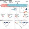
a | The clonal evolution of a neoplasm, during which four longitudinal samples (dotted lines) are taken, including samples from diagnostic biopsy, post-treatment biopsy, metastasectomy and resection of a late-emerging metastasis. The approximate timing of initiation of therapy, disease progression and death (autopsy) are also shown, and samples taken during autopsy are indicated by open arrowheads. Following the diagnostic biopsy, each subsequent sample increases the resolution of phylogenetic analyses. The caveat of this approach is that the interpretation is biased by the samples used. For example, the lack of a second sample of the primary tumour or from metastasis 2 limits inferences of a late subclonal event (dark blue clone), b | On the basis of multiregion sampling at autopsy, the phylogenetic analyses are more reflective of the dominant lethal subclone (dark blue) that emerged after two failed therapies but fail to capture the timing of emergence of subclones that were detected by longitudinal sampling (for example, in metastasis 1). c | Combination of the samples obtained by temporal sampling and multiregion sampling at autopsy reveal the complete evolutionary history of the neoplasm, illustrating that the combination of these approaches can yield more robust inferences of the clonal dynamics of the neoplasm.
Fig. 4 |
Fig. 4 |. Incorporation of research autopsies into biomarker-driven adaptive clinical trials.
This hypothetical biomarker-driven adaptive clinical trial begins with randomization to four drug regimens (arms 1–4). Patients who progress while receiving treatment are offered a research autopsy, in which a subset of patients will elect to participate. Interim analyses are performed with the goal of identifying the regimens that might be the most successful on the basis of treatment responses, and could be improved by using research autopsies to identify mechanisms and biomarkers of treatment resistance and progression. Such data from autopsies would also be expected to inform eligibility criteria for prospectively accrued patients.
Fig. 5 |
Fig. 5 |. Integration of multimodal data to maximize understanding of lethal cancer.
Many important initiatives have focused on one type of analysis in a large cohort of patients,. By contrast, research autopsies generate ample amounts of tissue to enable all types of analyses to be performed within the same patient, and even in the same piece of tissue. Thus, the scale of data possible from research autopsies requires computational efforts and innovation to maximize the use of this information and reveal biological aspects of lethal cancer that were not previously appreciated.

Similar articles

Cited by

References

    1. Buja LM, Barth RF, Krueger GR, Brodsky SV & Hunter RL The importance of the autopsy in medicine: perspectives of pathology colleagues. Acad. Pathol 6, 2374289519834041 (2019). - PMC - PubMed
    1. Blokker BM et al. Conventional autopsy versus minimally invasive autopsy with postmortem MRI, CT, and CT-guided biopsy: comparison of diagnostic performance. Radiology 289, 658–667 (2018). - PubMed
    1. Kretzschmar H Brain banking: opportunities, challenges and meaning for the future. Nat Rev. Neurosci 10, 70–78 (2009). - PubMed
    1. Hajdu SI A note from history: the first printed case reports of cancer. Cancer 116, 2493–2498 (2010). - PubMed
    1. Mariette C et al. Consensus on the pathological definition and classification of poorly cohesive gastric carcinoma. Gastric Cancer 22, 1–9 (2019). - PubMed

Publication types

Substances