

Anterior cruciate ligament grafts display differential maturation patterns on magnetic resonance imaging following reconstruction: a systematic review
- PMID: 31520146
- PMCID: PMC7067650
- DOI: 10.1007/s00167-019-05685-y
Anterior cruciate ligament grafts display differential maturation patterns on magnetic resonance imaging following reconstruction: a systematic review
Abstract
Purpose: The appearance of anterior cruciate ligament (ACL) grafts on magnetic resonance imaging (MRI) is related to graft maturity and mechanical strength after ACL reconstruction (ACLR). Accordingly, the purpose of this review was to quantitatively analyze reports of serial MRI of the ACL graft during the first year following ACLR; the hypothesis tested was that normalized MRI signal intensity would differ significantly by ACL graft type, graft source, and postoperative time.
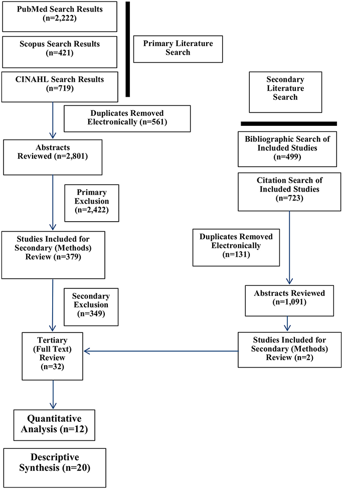
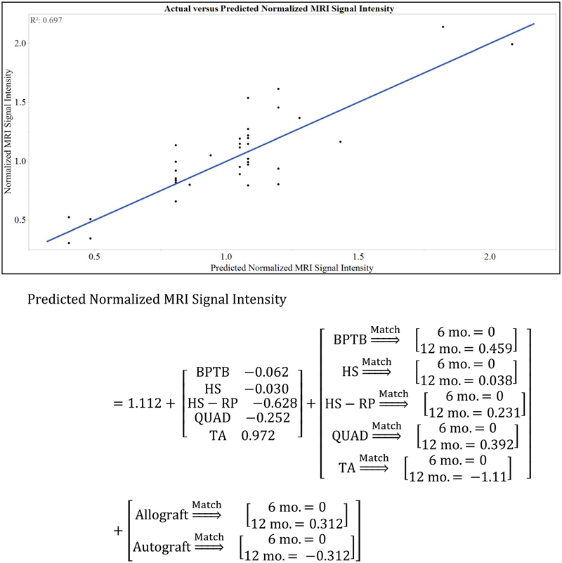
Methods: PubMed, Scopus, and CINAHL were searched for all studies published prior to June 2018 reporting MRI signal intensity of the ACL graft at multiple time points during the first postoperative year after ACLR. Signal intensity values at 6 and 12 months post-ACLR were normalized to initial measurements and analyzed using a least-squares regression model to study the independent variables of postoperative time, graft type, and graft source on the normalized MRI signal intensity.
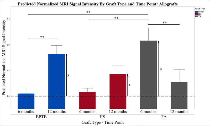
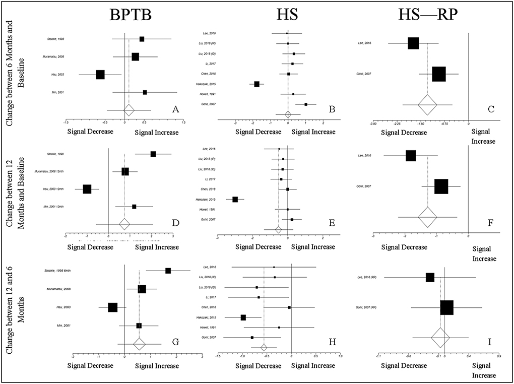
Results: An effect of graft type (P = 0.001) with interactions of graft type * time (P = 0.012) and graft source * time (P = 0.001) were observed. Post hoc analyses revealed greater predicted normalized MRI signal intensity of patellar tendon autografts than both hamstring (P = 0.008) and hamstring with remnant preservation (P = 0.001) autografts at postoperative month 12.
Conclusion: MRI signal varies with graft type, graft source, and time after ACLR. Enhanced graft maturity during the first postoperative year was associated with hamstring autografts, with and without remnant preservation. Serial MRI imaging during the first postoperative year may be clinically useful to identify biologically or mechanically deficient ACL grafts at risk for failure.
Level of evidence: IV.
Keywords: Anterior cruciate ligament; Ligamentization; Magnetic resonance imaging; Signal–noise-quotient.
Conflict of interest statement
Figures

Similar articles
-
Increased global posterior tibial slope is significantly associated with higher ACL graft signal intensity on 2-Year postoperative MRI after primary ACL reconstruction using hamstring tendon autografts.BMC Musculoskelet Disord. 2024 Nov 13;25(1):905. doi: 10.1186/s12891-024-08032-6. BMC Musculoskelet Disord. 2024. PMID: 39538228 Free PMC article.
-
Effect of Modified Lemaire Anterolateral Extra-articular Tenodesis on the Magnetic Resonance Imaging Maturity Signal of Anterior Cruciate Ligament Hamstring Graft.Am J Sports Med. 2021 Jul;49(9):2379-2386. doi: 10.1177/03635465211018858. Epub 2021 Jun 16. Am J Sports Med. 2021. PMID: 34133234
-
A Randomized Clinical Trial to Evaluate Attached Hamstring Anterior Cruciate Ligament Graft Maturity With Magnetic Resonance Imaging.Am J Sports Med. 2018 Apr;46(5):1143-1149. doi: 10.1177/0363546517752918. Epub 2018 Feb 14. Am J Sports Med. 2018. PMID: 29443537 Clinical Trial.
-
Anterior Cruciate Ligament Reconstruction: A Systematic Review and Meta-analysis of Outcomes for Quadriceps Tendon Autograft Versus Bone-Patellar Tendon-Bone and Hamstring-Tendon Autografts.Am J Sports Med. 2019 Dec;47(14):3531-3540. doi: 10.1177/0363546518825340. Epub 2019 Feb 21. Am J Sports Med. 2019. PMID: 30790526
-
Can Preoperative Magnetic Resonance Imaging Predict Intraoperative Autograft Size for Anterior Cruciate Ligament Reconstruction? A Systematic Review.J Knee Surg. 2019 Jul;32(7):649-658. doi: 10.1055/s-0038-1666830. Epub 2018 Jul 6. J Knee Surg. 2019. PMID: 29980152
Cited by
-
Why Should Return to Sport Be Delayed by up to Two Years After ACL Reconstruction? A Narrative Review of the Biological, Surgical and Rehabilitation Evidence.J Clin Med. 2025 Aug 12;14(16):5699. doi: 10.3390/jcm14165699. J Clin Med. 2025. PMID: 40869524 Free PMC article. Review.
-
Periosteal wrapping of the hamstring tendon autograft improves graft healing and prevents tunnel widening after anterior cruciate ligament anatomic reconstruction.Arch Orthop Trauma Surg. 2024 Jun;144(6):2711-2722. doi: 10.1007/s00402-024-05356-9. Epub 2024 May 15. Arch Orthop Trauma Surg. 2024. PMID: 38748257 Free PMC article.
-
Anterior cruciate ligament autograft maturation on sequential postoperative MRI is not correlated with clinical outcome and anterior knee stability.Knee Surg Sports Traumatol Arthrosc. 2022 Oct;30(10):3258-3267. doi: 10.1007/s00167-021-06777-4. Epub 2021 Nov 5. Knee Surg Sports Traumatol Arthrosc. 2022. PMID: 34739559 Free PMC article.
-
Extracorporeal Shockwave Therapy Improves Outcome after Primary Anterior Cruciate Ligament Reconstruction with Hamstring Tendons.J Clin Med. 2023 May 9;12(10):3350. doi: 10.3390/jcm12103350. J Clin Med. 2023. PMID: 37240456 Free PMC article.
-
A Comprehensive Framework to Evaluate the Effects of Anterior Cruciate Ligament Injury and Reconstruction on Graft and Cartilage Status through the Analysis of MRI T2 Relaxation Time and Knee Laxity: A Pilot Study.Life (Basel). 2021 Dec 10;11(12):1383. doi: 10.3390/life11121383. Life (Basel). 2021. PMID: 34947914 Free PMC article.
References
-
- Abe S, Kurosaka M, Iguchi T, Yoshiya S, Hirohata K (1993) Light and electron microscopic study of remodeling and maturation process in autogenous graft for anterior cruciate ligament reconstruction. Arthroscopy 9:394–405 - PubMed
-
- Ahn JH, Jeong HJ, Lee YS, Park JH, Lee JH, Ko TS (2016) Graft bending angle is correlated with femoral intraosseous graft signal intensity in anterior cruciate ligament reconstruction using the outside-in technique. Knee 23:666–673 - PubMed
-
- Ahn JH, Lee SH, Choi SH, Lim TK (2010) Magnetic resonance imaging evaluation of anterior cruciate ligament reconstruction using quadrupled hamstring tendon autografts: comparison of remnant bundle preservation and standard technique. Am J Sports Med 38:1768–1777 - PubMed
-
- Ahn JH, Lee YS, Jeong HJ, Park JH, Cho Y, Kim KJ et al. (2017) Comparison of transtibial and retrograde outside-in techniques of anterior cruciate ligament reconstruction in terms of graft nature and clinical outcomes: a case control study using 3T MRI. Arch Orthop Trauma Surg 137:357–365 - PubMed
-
- Amiel D, Kleiner JB, Roux RD, Harwood FL, Akeson WH (1986) The phenomenon of “ligamentization”: anterior cruciate ligament reconstruction with autogenous patellar tendon. J Orthop Res 4:162–172 - PubMed
Publication types
MeSH terms
Grants and funding
LinkOut - more resources
Full Text Sources
Medical
Research Materials
