. 2019 Dec;34(6):245-253.
doi: 10.1111/omi.12270. Epub 2019 Oct 21.

Type I interferon and interferon-stimulated gene expression in oral epithelial cells

Affiliations

Type I interferon and interferon-stimulated gene expression in oral epithelial cells

D C Brice et al. Mol Oral Microbiol. 2019 Dec.

Abstract

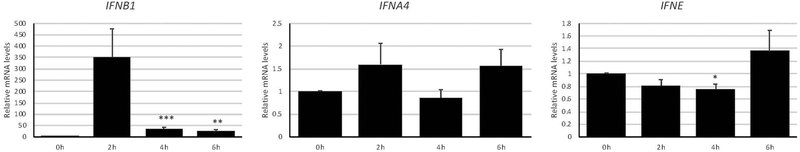
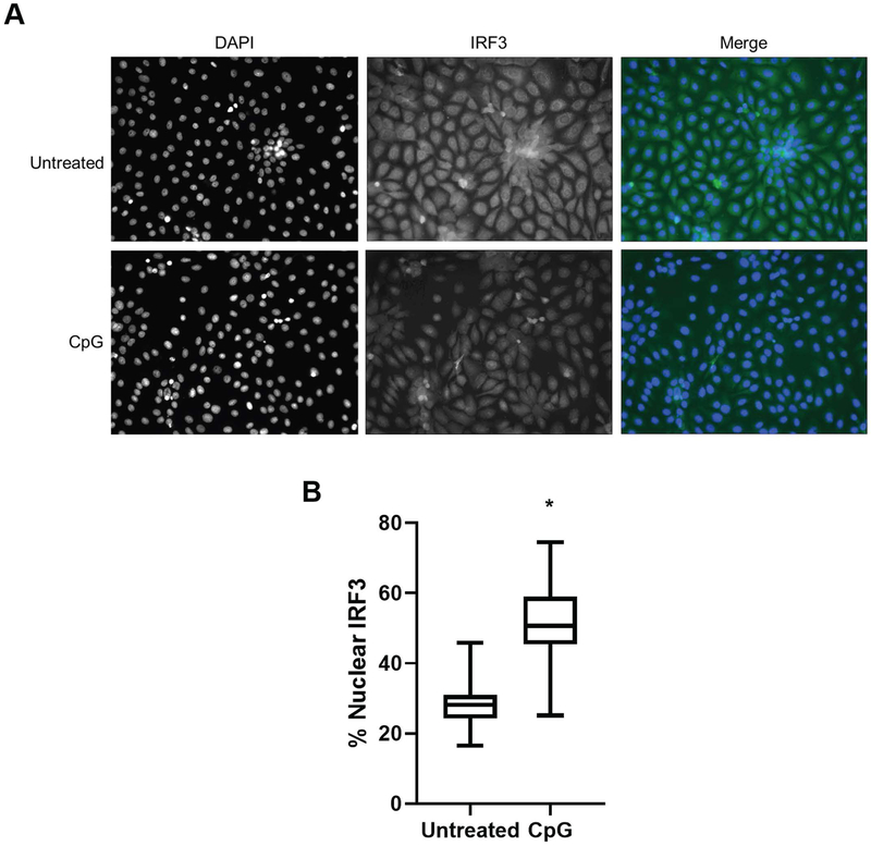
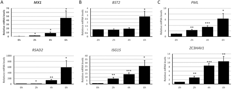
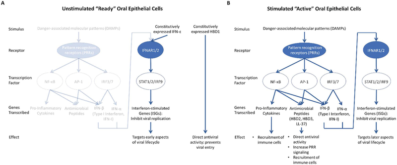
Oral epithelial cells (OEC) represent the first site of host interaction with viruses that infect the body through the oral route; however, their innate antiviral defense mechanisms yet to be defined. Previous studies have determined that OEC express pathogen-, damage-, or danger-associated molecular patterns (PAMPs or DAMPs), but their expression of key antiviral innate immune mediators, including type I interferons (type I IFN) and interferon-stimulated genes (ISGs) has not been studied extensively. We used the oral keratinocyte cell line, OKF6/TERT1, in the presence and absence of the viral mimics poly(I:C) and unmethylated CpG DNA, to define the expression of type I IFN and ISGs. We identified the basal expression of novel type I IFN genes IFNE and IFNK, while IFNB1 was induced by viral mimics, through the nuclear translocation of IRF3. Numerous ISGs were expressed at basal levels in OEC, with an apparent correlation between high expression and antiviral activity at the earlier stages of viral infection. Stimulation of OECs with poly(I:C) led to selective induction of ISGs, including MX1, BST2, PML, RSAD2, ISG15, and ZC3HAV1. Together, our results demonstrate that OECs exhibit a robust innate antiviral immune defense profile, which is primed to address a wide variety of pathogenic viruses that are transmitted orally.

Keywords: epithelial; innate immunity; interferon; interferon-stimulated gene; oral; virus.


Conflict of interest statement

The authors have no conflicts of interest to declare.

Figures

Figure 1.
Figure 1.. Type I interferon expression in oral epithelial cells upon pattern recognition receptor stimulation.
OKF6/Tert-1 cells were treated with 1 μg/ml poly(I:C) for up to six hours and measured for changes in type I interferon expression by RT-qPCR. Bar graphs and error bars represent the means and standard errors of measurement (SEM) of three independent experiments, each with three biological replicates per condition. *p < 0.05, **p < 0.005, and ***p < 0.0005 compared to 0h, using a two-tailed, unpaired Student’s t-test. All data analysis was performed using GraphPad Prism version 8.2.0 for Windows, GraphPad Software, La Jolla California USA, www.graphpad.com.
Figure 2.
Figure 2.. Oral epithelial cell IRF3 localization upon pattern recognition receptor stimulation.
OKF6/Tert-1 cells were treated with with 5 μM unmethylated CpG DNA for two hours. Localization of IRF3, either within or outside the nucleus as determined by co-localization with DAPI staining, was determined by immunofluoresence microscopy (A). Percentage of IRF3 localized to the nuclei of cells was measured for fifty cells per condition (B). *p < 0.0001 compared to the untreated condition, a two-tailed, unpaired Student’s t-test after determining data from both conditions were normally distributed using the Shapiro-Wilk test.
Figure 3.
Figure 3.. Interferon-stimulated gene expression in oral epithelial cells upon pattern recognition receptor stimulation.
OKF6/Tert-1 cells were treated with 1 μg/ml poly(I:C) for up to six hours and measured for changes in interferon-stimulated gene (ISG) expression by RT-qPCR. Measured genes were divided into each ISG basal expression group, according to Table 2: Low (A), Medium (B), and High (C). Bar graphs and error bars represent the means and standard errors of measurement (SEM) of three independent experiments, each with three biological replicates per condition. *p < 0.05, **p < 0.005, and ***p < 0.0005 compared to 0h, using a two-tailed, unpaired Student’s t-test.
Figure 4.
Figure 4.. Proposed model of oral epithelial cell innate antiviral defenses.
Oral epithelial cells (OECs) either exist as unstimulated and “ready” for a potential viral infection (A) or stimulated and “active” in the defense against a replicating virus (B). (A) “Ready” OECs are not stimulated by danger-associated molecular patterns (DAMPs) and, therefore, do not express pro-inflammatory cytokines. However, despite not expressing the full repertoire of either type I interferon (type I IFN) or antimicrobial peptides (also known as host defense peptides, HDPs), OECs do express some of these proteins constitutively, leading to direct antiviral activity mediated by human beta defensin 1 (HBD1) (Brice & Diamond, 2019; Zhao, Wang, & Lehrer, 1996) and certain interferon-stimulated genes (ISGs) that target early aspects of the viral lifecycle. (B) OECs become “active” when stimulated with DAMPs, leading to transcription of a larger number of pro-inflammatory cytokines, HDPs, and type I IFN, each with their own antiviral effects.

References

    1. Akira S, Uematsu S, & Takeuchi O (2006). Pathogen recognition and innate immunity. Cell, 124(4), 783–801. 10.1016/j.cell.2006.02.015 - DOI - PubMed
    1. Brice DC, & Diamond G (2019). Antiviral Activities of Human Host Defense Peptides. Current Medicinal Chemistry, 26 10.2174/0929867326666190805151654 - DOI - PMC - PubMed
    1. Brice DC, Toth Z, & Diamond G (2018). LL-37 disrupts the Kaposi’s sarcoma-associated herpesvirus envelope and inhibits infection in oral epithelial cells. Antiviral Research, 158, 25–33. 10.1016/J.ANTIVIRAL.2018.07.025 - DOI - PMC - PubMed
    1. Brubaker SW, Bonham KS, Zanoni I, & Kagan JC (2015). Innate immune pattern recognition: a cell biological perspective. Annual Review of Immunology, 33, 257–290. 10.1146/annurev-immunol-032414-112240 - DOI - PMC - PubMed
    1. Cervantes CAC, Oliveira LMS, Manfrere KCG, Lima JF, Pereira NZ, Duarte AJS, & Sato MN (2016). Antiviral factors and type I/III interferon expression associated with regulatory factors in the oral epithelial cells from HIV-1-serodiscordant couples. Scientific Reports, 6(1), 25875 10.1038/srep25875 - DOI - PMC - PubMed

Publication types