. 2019 Nov:188:107798.
doi: 10.1016/j.exer.2019.107798. Epub 2019 Sep 11.

Novel anti-angiogenic PEDF-derived small peptides mitigate choroidal neovascularization

Affiliations

Novel anti-angiogenic PEDF-derived small peptides mitigate choroidal neovascularization

Nader Sheibani et al. Exp Eye Res. 2019 Nov.

Abstract

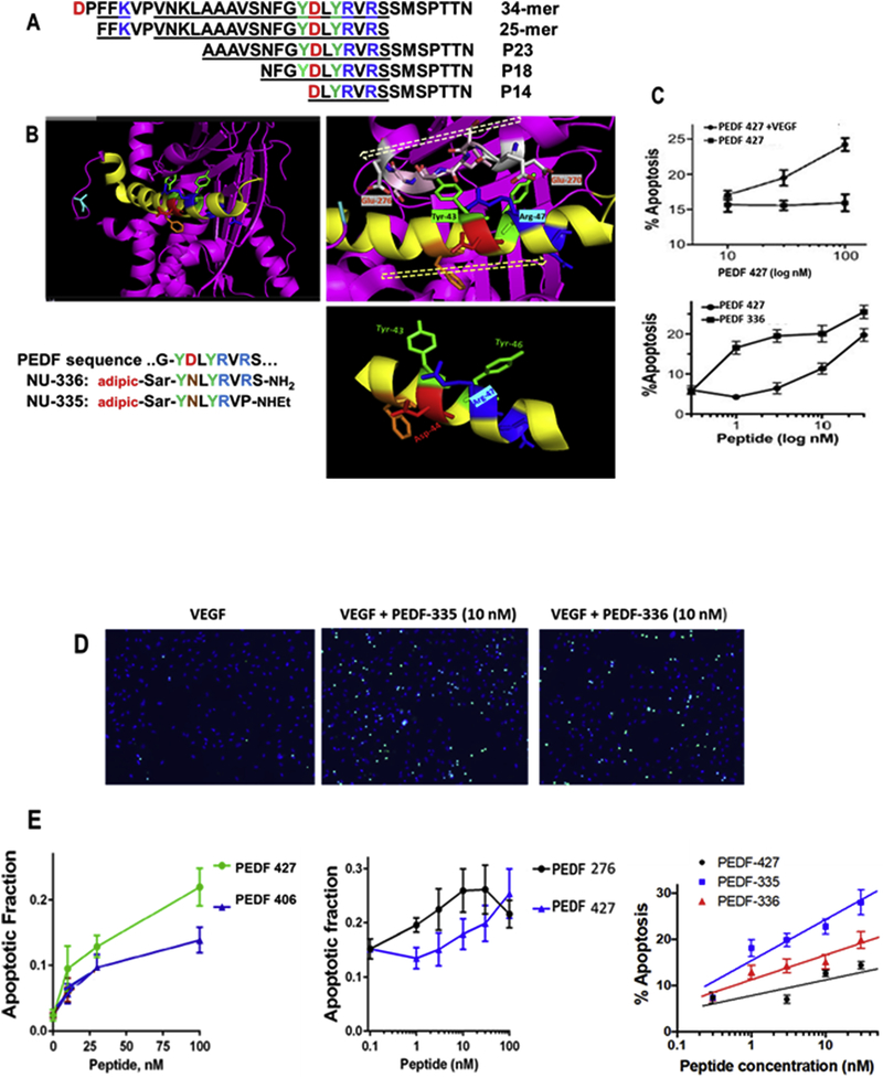
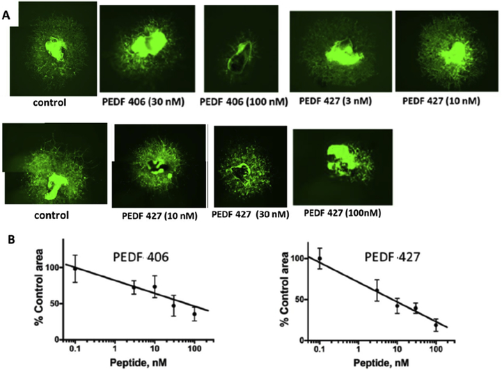
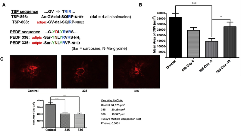
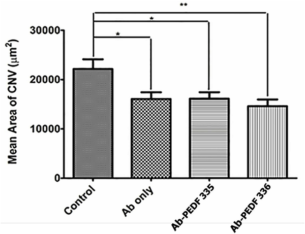
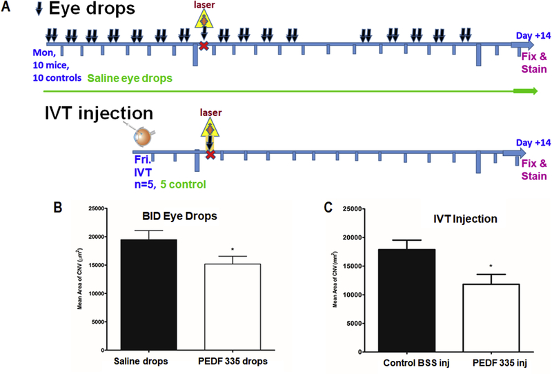
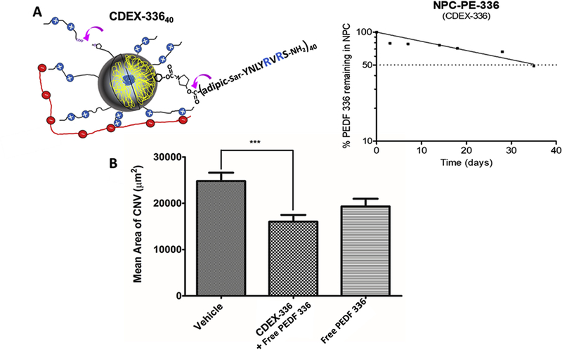
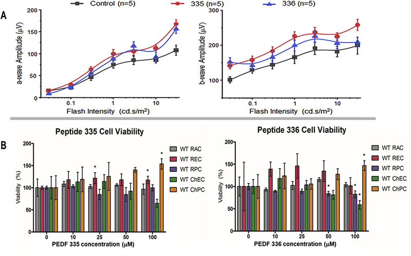
Abnormal migration and proliferation of endothelial cells (EC) drive neovascular retinopathies. While anti-VEGF treatment slows progression, pathology is often supported by decrease in intraocular pigment epithelium-derived factor (PEDF), an endogenous inhibitor of angiogenesis. A surface helical 34-mer peptide of PEDF, comprising this activity, is efficacious in animal models of neovascular retina disease but remains impractically large for therapeutic use. We sought smaller fragments within this sequence that mitigate choroidal neovascularization (CNV). Expecting rapid intravitreal (IVT) clearance, we also developed a method to reversibly attach peptides to nano-carriers for extended delivery. Synthetic fragments of 34-mer yielded smaller anti-angiogenic peptides, and N-terminal capping with dicarboxylic acids did not diminish activity. Charge restoration via substitution of an internal aspartate by asparagine improved potency, achieving low nM apoptotic response in VEGF-activated EC. Two optimized peptides (PEDF 335, 8-mer and PEDF 336, 9-mer) were tested in a mouse model of laser-induced CNV. IVT injection of either peptide, 2-5 days before laser treatment, gave significant CNV decrease at day +14 post laser treatment. The 8-mer also decreased CNV, when administered as eye drops. Also examined was a nanoparticle-conjugate (NPC) prodrug of the 9-mer, having positive zeta potential, expected to display longer intraocular residence. This NPC showed extended efficacy, even when injected 14 days before laser treatment. Neither inflammatory cells nor other histopathologic abnormalities were seen in rabbit eyes harvested 14 days following IVT injection of PEDF 336 (>200 μg). No rabbit or mouse eye irritation was observed over 12-17 days of PEDF 335 eye drops (10 mM). Viability was unaffected in 3 retinal and 2 choroidal cell types by PEDF 335 up to 100 μM, PEDF 336 (100 μM) gave slight growth inhibition only in choroidal EC. A small anti-angiogenic PEDF epitope (G-Y-D-L-Y-R-V) was identified, variants (adipic-Sar-Y-N-L-Y-R-V) mitigate CNV, with clinical potential in treating neovascular retinopathy. Their shared active motif, Y - - - R, is found in laminin (Ln) peptide YIGSR, which binds Ln receptor 67LR, a known high-affinity ligand of PEDF 34-mer.

Keywords: Angiogenesis; Choroidal neovascularization; Laminin receptor; Macular degeneration; PEDF; Peptides; Retinopathy; YIGSR.


Figures

Figure 1.
Figure 1.. Secondary structure and mimicry of PEDF N-terminal 34-mer peptide.
(A) The active anti-angiogenic, largely α-helical 34-mer extends from Asp-24 to Asn-56 of PEDF (Filleur et al., 2005). An active N-terminal 25-mer therein bound tightly to 67LR (Bernard et al., 2009), and more distal 23-mer and 18-mer (P18) fragments have also been shown active, although a 14-mer starting at Asp44 was inactive (Mirochnik et al., 2009). Amino acid sequences are shown with positively charged residues in blue, negative residues red and Tyr in green, with shaded alpha-helical span from Asn32 through Ser52. (B) The PEDF x-ray crystal structure Protein Data Base PDB ID 1IMV, (Simonovic et al., 2001) is shown as ribbon backbone, the above region highlighted yellow. It is then enlarged from N32-S52, hashed arrow left-to-right tracing N-to-C direction. This reveals Arg47 accessible to solvent, with Tyr43 partially overlaid by residues 270–276 (EESKLTSE, hashed arrow right-to-left N-to-C direction) in a disordered segment, likely able to move, allowing Tyr43 solvent access. Bottom right panel is the Asn39 to Met51 portion where side chains of Tyr43 and Arg47 are seen on the same helical face, replicated in synthetic peptides shown. (C) Apoptosis assay, as described in section 2.7, shows VEGF requirement, also peptide concentration dependence indicates PEDF 336 improvement over PEDF 427. D) EC apoptosis was detected by in situ TUNEL (green), cell nuclei highligfhted by DAPI (blue) with representative images of EC apoptosis shown. Quantitative analysis performed using Nikon Elements software and percent apoptotic cells calculated for each treatment. A minimum of 8 field per condition (100–300 cells per field) were analyzed. (E) Linear regression plots were generated using Prizm 6 software package. Pairwise and 3-peptide simultaneous comparisons are shown as concentration dependence, leading to the potency ranking in Table 1. Statistical significance was determined using multiple T test (P<0.0001).
Figure 2.
Figure 2.. PEDF peptides inhibit choroidal neovascularization ex vivo.
Choroidal explants were generated from eyes harvested from C57Bl6 mice that carry eGFP transgene under beta-actin promoter. The explants were embedded in Matrigel drops and cultured in media containing VEGF alone or with increasing concentrations of selected PEDF peptides from Table 1. The explants were observed daily and images were acquired at 6 days endpoint. Composite images were generated for larger vascular areas to evaluate the full vascular area. (A) Representative images of VEGF-stimulated choroidal explants treated with PEDF 406 at increasing concentrations. Similar images were obtained for PEDF 427. (B) Quantitative analysis of the images as shown in A. Sprouting area was measured using ImageJ software and the area of explant subtracted for each image. A minimum of 3 explants were analyzed per data point. Statistical significance was determined using multiple T-test and P value is below 0.0001 for both C and D.
Figure 3.
Figure 3.. Peptide inhibition of CNV.
(A) TSP1-derived and PEDF-derived test peptide sequences are shown in relation to original protein sequence. Benchmark anti-angiogenic TSP-1 mimetic peptide (ABT-898): Ac-GV-d-alloIle-Ser-Q-I-R-Pro-ethylamide has been shown to inhibit laser-induced mouse CNV (Wang et al., 2012). Substituting N-terminal adipic acid as a half-amide in place of the N-acetyl group gave TSP-868, also displaying anti-CNV activity, with potential capacity to form an adipic half-ester prodrug. (B) TSP-868 (2μL, 2 mM) was compared against balanced salts vehicle at different times of IVT injection where day 0 is the day of laser treatment. CNV mitigation by peptide TSP-868 was significant when 4 ng in 2 μL PBS was IVT-injected on the day of laser, with effect enhanced by injecting at day −2 (not shown) or day −5 before laser treatment. No isignificant inhibition of CNV was evident with injection at day −10. (C) Peptides PEDF 335 and PEDF 336 were IVT injected (2μL, 2 mM) 2 days prior to laser treatment. PEDF-335 and PEDF-336 IVT injection at day −2 significantly decreased CNV by 40–50%, similar inhibition was obtained with injection on day −5 (not shown). In all cases eyes were harvested for CNV analysis at day +14.
Figure 4.
Figure 4.. Evaluation of anti-VEGF alone or in combination with PEDF peptides.
Mice were IVT- injected with anti-mouse VEGF164 (1 μL of 25 μg/mL alone or in combination with PEDF peptides (1 μL 4 mM peptide in the antibody solution) on day −2. The vehicle was Avastin-vehicle per package insert (trehalose, phosphate, NaCl), and was used for preparing all solutions. Animals were sacrificed on day 14 and RPE/choroidal flatmounts were prepared and stained with anti-ICAM-2 as described under “Methods”.The lesion areas were determined using imageJ. Please note a significant inhibition of neovascularization with antibody alone (*P< 0.05) and similar response with antibody and PEDF-335 (*P< 0.05). Statistical significance was enhanced by PEDF-336 (**P< 0.01).
Figure 5.
Figure 5.. PEDF 335 delivery via eye drops was effective in attenuation of CNV.
Animals (5 per group) received vehical alone or PEDF 335 peptide by one-time IVT injection (1 μL of 4 mM peptide). The topical treatment groups (10 mice / group received 10 mM peptide or saline (5 μL drop, bid, Mon-Fri) starting at day −7. (A) shows the schedule of eye drop or IVT dosing and eye harvest and fixation. Animals were then treated with laser on day 0 and topical delivery of the peptide or saline control drops continued Mon-Fri during the ensuing 14 day period. Animals were then sacrificed and RPE/Choroid flatmounts were stained with ICAM-2 antibody and was determined as described in Methods. (B) Area of vascularization for IVT injection at day −3. (C) Area of vascularization for topical treatment. Inhibition was similarly significant for both treatments, (*P< 0.05).
Figure 6.
Figure 6.. CDEX NPC peptide ester prodrug extends anti-CNV effect.
(A) Pyrrolydyl-3OH ester of PEDF 336 (PE-336), was carbamate-conjugated to glucose OH groups in CDEX, giving prodrug NPC-PE-(336)40, with ζ = +2.3 mV (Table 2). (A) Representation at left shows NPC prodrug adherence to IVT HA by multivalent ionic interaction (Melgar-Asensio et al., 2018), purple arrows point to sites of spontaneous ester hydrolysis. Released peptide at 154 days (taken as 95% completion of hydrolysis) and at earlier incubation times was measured via UV spectra of ultrafiltrates. Un-hydrolyzed prodrug ester covalently attached to NPC at these times was: % remaining = 100–100 × (0.295 - OD275nm ultrafiltrate)/(0.295), with calculated release t1/2 of 35 +/− 5 days based on one phase exponential decay analysis by GraphPad Prism version 5, GraphPad Software, La Jolla, CA. Semi-log loss of free PEDF 336 is shown on the right under physiological conditions in buffer. (B) IVT injection was carried out 14 days prior to laser induction. For free peptide this was 2 μL containing 4 nmoles of PEDF 336. CDEX-336 injection was 2 μL containing 2 nmoles of free peptide and 1.3 nmoles (1.5 μg) of peptide as prodrug in (4.3 μg NPC), expected daily peptide release approximately 20–30 ng. CNV was estimated as described at 28 days post-injection, only the NPC prodrug mixture showed statistically significant CNV reduction (**P< 0.01).
Figure 7.
Figure 7.. Peptide safety: ERG and cytotoxicity.
(A) Average a- (left panel) and b-wave (right panel) amplitudes are shown in response to increasing flash intensities for mice ( 5 per group, 10 eyes) after injected with 2 μL of 335 (red circle), 336 (blue triangle) peptide or BSS (black square, balanced salts solution-vehicle control). Data represented as average ± SEM. (B) Cell viability in response to peptide treatments. Various cell types were incubated with indicated concentration of peptides and cell viability was assessed after 48 h using the Cell Titer 96® Non-Radioactive Cell Proliferation Assay (MTT) as recommended by the supplier. The data is normalized to control treated cells and shows mean ± SEM from three independent observations. Abbreviations: RAC, retinal astrocytes; REC, retinal endothelial cells; RPC, retinal pericytes; ChEC, choroidal endothelial cells; ChPC, choropidal pericytes.

References

    1. Alsenbrey S, Zhang M, Bacher D, Yee J, Brunken WJ, Hunter DD, 2006. Retinal pigment epithelial cells synthesize laminins, including laminin 5, and adhere to them through alpha3- and alpha6-containing integrins. Invest Ophthalmol Vis Sci. 47 (12),5537–5544. 10.1167/iovs.05-1590 - DOI - PMC - PubMed
    1. Amaral J, Becerra SP, 2010. Effects of human recombinant PEDF protein and PEDF-derived peptide 34-mer on choroidal neovascularization. Invest Ophthalmol Vis Sci. 51 (3), 1318–1326. 10.1167/iovs.09-4455 - DOI - PMC - PubMed
    1. Ardini E, Sporchia B, Pollegioni L, Modugno M,Ghirelli C, Castiglioni F,Tagliabue E, Menard S, 2002. Identification of a novel function for 67-kDa laminin receptor: increase in laminin degradation rate and release of motility fragments. Canc Res. 62, 1321–1325. https://www.ncbi.nlm.nih.gov/pubmed/11888899 - PubMed
    1. Askou A,L, Alsing S, Benckendorff JNE, Holmgaard A, Jacob Giehm Mikkelsen JG, Aagaard L, Bek T, Thomas J, Corydon TJ 2019. Suppression of choroidal neovascularization by AAV-based dual-Acting antiangiogenic gene therapy. Mol Ther Nucleic Acids. 16, 38–50. 10.1016/j.omtn.2019.01.012 - DOI - PMC - PubMed
    1. Austin A, Lietman T, Rose-Nussbaumer J, 2017. Update on the management of infectious keratitis. Ophthalmol.124 (11), 1678–1616. 10.1016/j.ophtha.2017.05.012 - DOI - PMC - PubMed

Publication types

MeSH terms