Abbreviated quantitative UTE imaging in anterior cruciate ligament reconstruction
- PMID: 31521135
- PMCID: PMC6745079
- DOI: 10.1186/s12891-019-2811-x
Abbreviated quantitative UTE imaging in anterior cruciate ligament reconstruction
Abstract
Background: Existing ultrashort echo time magnetic resonance imaging (UTE MRI) methods require prohibitively long acquisition times (~ 20-40 min) to quantitatively assess the clinically relevant fast decay T2* component in ligaments and tendons. The purpose of this study was to evaluate the feasibility and clinical translatability of a novel abbreviated quantitative UTE MRI paradigm for monitoring graft remodeling after anterior cruciate ligament (ACL) reconstruction.
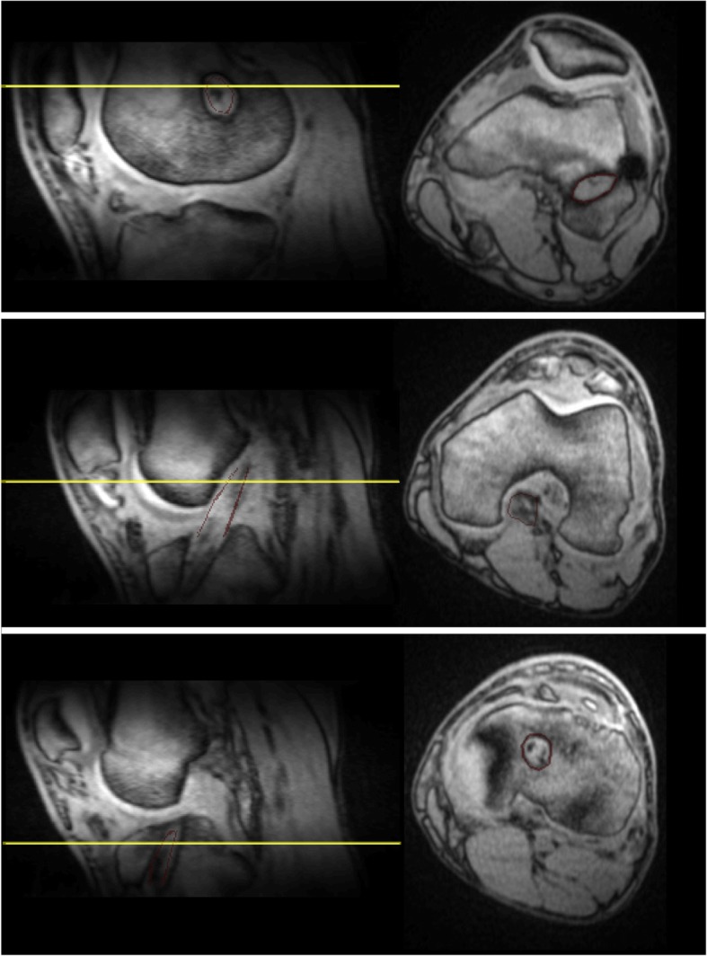
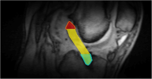
Methods: Eight patients who had Graftlink™ hamstring autograft reconstruction were recruited for this prospective study. A 3D double-echo UTE sequence at 3.0 Tesla was performed at 3- and 6-months post-surgery. An abbreviated UTE MRI paradigm was established based on numerical simulations and in vivo validation from healthy knees. This proposed approach was used to assess the T2* for fast decay component ([Formula: see text]) and bound water signal fraction (fbw) of ACL graft in regions of interest drawn by a radiologist.
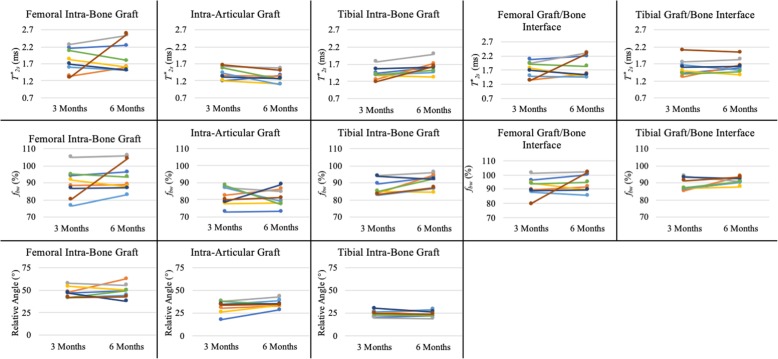
Results: Compared to the conventional bi-exponential model, the abbreviated UTE MRI paradigm achieved low relative estimation bias for [Formula: see text] and fbw over a range of clinically relevant values for ACL grafts. A decrease in [Formula: see text] of the intra-articular graft was observed in 7 of the 8 ACL reconstruction patients from 3- to 6-months (- 0.11 ± 0.16 ms, P = 0.10). Increases in [Formula: see text] and fbw from 3- to 6-months were observed in the tibial intra-bone graft ([Formula: see text]: 0.19 ± 0.18 ms, P < 0.05; Δfbw: 4% ± 4%, P < 0.05). Lower [Formula: see text] (- 0.09 ± 0.11 ms, P < 0.05) was observed at 3-months when comparing the intra-bone graft to the graft/bone interface in the femoral tunnel. The same comparisons at the 6-months also yielded relatively lower [Formula: see text] (- 0.09 ± 0.12 ms, P < 0.05).
Conclusion: The proposed abbreviated 3D UTE MRI paradigm is capable of assessing the ACL graft remodeling process in a clinically translatable acquisition time. Longitudinal changes in [Formula: see text] and fbw of the ACL graft were observed.
Keywords: ACL reconstruction; Anterior cruciate ligament (ACL); Graft healing; Ligamentization; Tendon-to-bone healing; Ultrashort TE (UTE).
Conflict of interest statement
Dr. James M. Paci is an educational consultant for and receives research support from Arthrex and Zimmer-Biomet.
Figures


Similar articles
-
Quantitative Assessment of In Vivo Human Anterior Cruciate Ligament Autograft Remodeling: A 3-Dimensional UTE-T2* Imaging Study.Am J Sports Med. 2020 Oct;48(12):2939-2947. doi: 10.1177/0363546520949855. Epub 2020 Sep 11. Am J Sports Med. 2020. PMID: 32915640
-
Graft position in arthroscopic anterior cruciate ligament reconstruction: anteromedial versus transtibial technique.Arch Orthop Trauma Surg. 2016 Nov;136(11):1571-1580. doi: 10.1007/s00402-016-2532-7. Epub 2016 Aug 2. Arch Orthop Trauma Surg. 2016. PMID: 27484876
-
Hamstring tendon autografts do not show complete graft maturity 6 months postoperatively after anterior cruciate ligament reconstruction.Knee Surg Sports Traumatol Arthrosc. 2019 Jan;27(1):130-136. doi: 10.1007/s00167-018-5033-0. Epub 2018 Jul 14. Knee Surg Sports Traumatol Arthrosc. 2019. PMID: 30008057 Free PMC article.
-
Anterior cruciate ligament grafts display differential maturation patterns on magnetic resonance imaging following reconstruction: a systematic review.Knee Surg Sports Traumatol Arthrosc. 2020 Jul;28(7):2124-2138. doi: 10.1007/s00167-019-05685-y. Epub 2019 Sep 13. Knee Surg Sports Traumatol Arthrosc. 2020. PMID: 31520146 Free PMC article.
-
A meta-analysis of bone-patellar tendon-bone autograft versus four-strand hamstring tendon autograft for anterior cruciate ligament reconstruction.Knee. 2015 Mar;22(2):100-10. doi: 10.1016/j.knee.2014.11.014. Epub 2014 Dec 11. Knee. 2015. PMID: 25547048 Review.
Cited by
-
Three-Dimensional Reconstruction Algorithm-Based Magnetic Resonance Imaging Evaluation of Biomechanical Changes in Articular Cartilage in Patients after Anterior Cruciate Ligament Reconstruction.Comput Intell Neurosci. 2022 Mar 15;2022:8256450. doi: 10.1155/2022/8256450. eCollection 2022. Comput Intell Neurosci. 2022. PMID: 35330602 Free PMC article.
-
Ultra-short T2 components imaging of the whole brain using 3D dual-echo UTE MRI with rosette k-space pattern.Magn Reson Med. 2023 Feb;89(2):508-521. doi: 10.1002/mrm.29451. Epub 2022 Sep 25. Magn Reson Med. 2023. PMID: 36161728 Free PMC article.
-
Association Between T2* Relaxation Times Derived From Ultrashort Echo Time MRI and Symptoms During Exercise Therapy for Patellar Tendinopathy: A Large Prospective Study.J Magn Reson Imaging. 2021 Nov;54(5):1596-1605. doi: 10.1002/jmri.27751. Epub 2021 May 30. J Magn Reson Imaging. 2021. PMID: 34056788 Free PMC article. Clinical Trial.
-
Reproducibility and postacquisition correction methods for quantitative magnetic resonance imaging of the anterior cruciate ligament (ACL).J Orthop Res. 2022 Dec;40(12):2908-2913. doi: 10.1002/jor.25319. Epub 2022 Mar 14. J Orthop Res. 2022. PMID: 35266588 Free PMC article.
-
Ligamentization of the reconstructed ACL differs between the intraarticular and intraosseous regions: A quantitative assessment using UTE-T2* mapping.PLoS One. 2022 Jul 22;17(7):e0271935. doi: 10.1371/journal.pone.0271935. eCollection 2022. PLoS One. 2022. PMID: 35867680 Free PMC article.
References
MeSH terms
LinkOut - more resources
Full Text Sources
Medical