. 2019 Sep 14;19(1):923.
doi: 10.1186/s12885-019-6130-2.

A way to understand idiopathic senescence and apoptosis in primary glioblastoma cells - possible approaches to circumvent these phenomena

Affiliations

A way to understand idiopathic senescence and apoptosis in primary glioblastoma cells - possible approaches to circumvent these phenomena

Karolina Janik et al. BMC Cancer. .

Abstract

Background: Glioblastoma (GB) is considered one of the most lethal tumors. Extensive research at the molecular level may enable to gain more profound insight into its biology and thus, facilitate development and testing of new therapeutic approaches. Unfortunately, stable glioblastoma cell lines do not reflect highly heterogeneous nature of this tumor, while its primary cultures are difficult to maintain in vitro. We previously reported that senescence is one of the major mechanisms responsible for primary GB cells stabilization failure, to a lesser extent accompanied by apoptosis and mitotic catastrophe-related cell death.

Methods: We made an attempt to circumvent difficulties with glioblastoma primary cultures by testing 3 different approaches aimed to prolong their in vitro maintenance, on a model of 10 patient-derived tumor specimens.

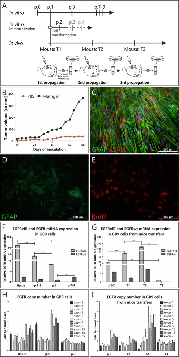
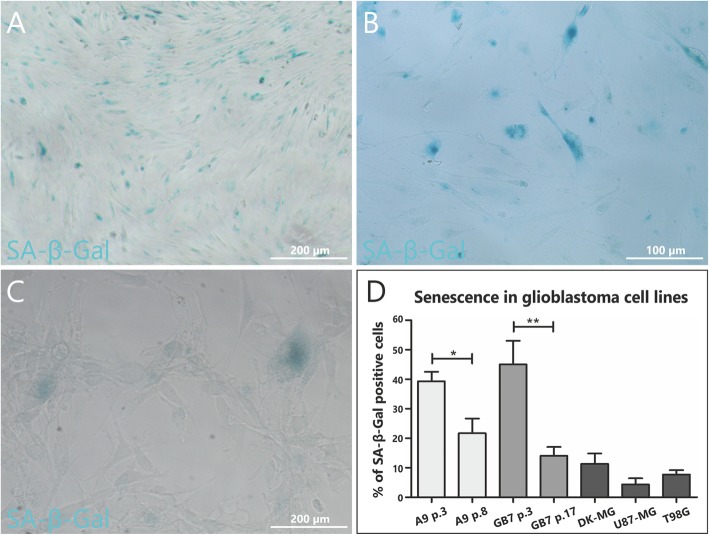
Results: Two out of ten analyzed GB specimens were successfully stabilized, regardless of culture approach applied. Importantly, cells transduced with immortalizing factors or cultured in neural stem cell-like conditions were still undergoing senescence/apoptosis. Sequential in vivo/in vitro cultivation turned out to be the most effective, however, it only enabled to propagate cells with preserved molecular profile up to 3rd mice transfer. Nevertheless, it was the only method that impeded these phenomena long enough to provide sufficient amount of material for in vitro/in vivo targeted analyses. Interestingly, our data additionally demonstrated that some subpopulations of several stabilized GB cell lines undergo idiopathic senescence, however, it is counterbalanced by simultaneous proliferation of other cell subpopulations.

Conclusions: In the majority of primary glioma cultures, there has to be an imbalance towards apoptosis and senescence, following few weeks of rapid proliferation. Our results indicate that it has to be associated with the mechanisms other than maintenance of glioblastoma stem cells or dependence on proteins controlling cell cycle.

Keywords: Apoptosis; Glioblastoma; Immortalization; In vivo model; Senescence.


Conflict of interest statement

The authors declare that they have no competing interests.

Figures

Fig. 1
Fig. 1
Monitoring of the stability of molecular alterations in GB cell cultures. a-c Representative figures showing results of conducted molecular analyses: FISH result presenting chromosome 7 trisomy and EGFR amplification in single cells of GB7 in passage 1 of monolayer culture (a); MLPA analysis showing CDKN2A deletion in GB5 in passage 3 of monolayer conditions (b); IDH1 sequencing of GB4 – the line marks the mutated nucleotide in codon 132 (R132H) (c); Changes in CDKN2A status (d), EGFRvIII and EGFRWT DNA copy number (e) and EGFRWT and EGFRvIII mRNA expression (f) in the culture course of glioblastoma cells in two analyzed culture conditions. EGFR probe (red signals); CEP 7 control probe – centromere of chromosome 7, enabling to show the number of chromosomes 7 (green signals). For MLPA results error bars indicate SD, while for Real-time PCR SEM. Statistical significance for Real-time PCR results was determined by two-way ANOVA analysis with post-analysis Bonferroni’s multiple comparisons test to compare each sample to the adequate frozen sample. Results were considered statistically significant at * p < 0.05; ** p < 0.01; *** p < 0.005
Fig. 2
Fig. 2
The comparison of NSC-like and classical monolayer conditions for culturing of primary glioblastoma cells. Both approaches did not enable to maintain long-term culture of GB cells and to avoid senescence/apoptosis; a-e Representative pictures showing results of the immunocytochemical analyses; a Early passage in monolayer serum conditions showing GFAP (+) GB cells; b Single GFAP (+), SA-β-Gal (+) GB cells from monolayer GB9 culture (intermediate passage) surrounded by cells negative for both these markers; c Early fresh GB9 neuropshere transferred onto poly-l-ornithine/laminin-coated dish, with the majority of GFAP (+) cells migrating from the neurosphere and only several αSMA (+) cells; d In late passage only large, flat and SA-β-Gal (+) cells with features of mitotic catastrophe maintained in culture; Inset boxes in B and D show higher magnifications of senescent (SA-β-Gal-positive) cells; e Intermediate passage of GB9 in NSC-like conditions – only GFAP (+) cells maintained in culture, but there is a lack of cells in mitosis; f Reduced proliferation rate of GB8 cells under NSC-like conditions vs. adherent monolayer culture; g-j The percentage of spontaneously senescent (as determined by means of SA-β-Gal assay), apoptotic (assessed using in vitro real-time caspase assay after 48 h of incubation with Caspase 3/7 synthetic reporter solution), proliferating (analyzed during 5 days incubation with BrdU), as well as other cells in various culture conditions and passages. The characteristics of cells in different culture conditions is depicted separately. The average percentage of cells with a particular feature was calculated by analyzing at least 200 cells per case from indicated passages of GB6-GB9 cultures. Error bars indicate SEM. k Statistical significance was calculated by paired Student’s t-test with p < 0.05 considered statistically significant
Fig. 3
Fig. 3
Spontaneous/idiopathic mitotic catastrophe (MC) in primary glioblastoma cultures. Polynucleated cell following MC in GB6 in early monolayer culture conditions (a) as well as abnormal mitoses in early passages of NSC-like conditions in GB9 are visible; (b-c) Multipolar spindles in GFAP positive cell abnormal mitosis (b) as well as polynuclear cells with asymmetric distribution of phosphorylated histone 3 may be easily detected in early passages of GB 7 in NSC-like conditions (c); The percentage of cells with the features of mitotic catastrophe (abnormal metaphases with spindle apparatus/chromosomal misalignments, mono- and multipolar spindles, bi- and multinuclear cells formed following MC) differs depending on case (GB6 and GB9) in the same conditions, here NSC-like ones; (d) Cells with the features of MC may be simultaneously SA-β-Gal positive or pass through mitotic cell death, as polynucleated senescent cell in GB9 in passage 4 of NSC-like conditions (e-f). The average percentage was obtained by analyzing of at least 200 cells per case (GB6 and GB9). Error bars indicate SEM
Fig. 4
Fig. 4
Primary glioblastoma cells transduced with immortalizing factors – none of applied factors successfully stabilized GB cell culture. a Table summarizing attempts to transduce GB cells at early culture stages with immortalizing factors; b Late passage of αSMA (+) pericytes from GB9 sample immortalized with lentivirus with SV40 + BMI1; c GB9 cells immortalized with lentivirus with BMI + hEST2 + SV40 and SV40 alone. Significant decrease in EGFRvIII and EGFRWT expression following immortalization attempt was observed; d-e Early passage of primary GB7 culture (mostly GFAP (+), SA-β-Gal (+) cells) treated with SV40 + BMI. ‘∞’ infinite proliferation; “F” cells did not get to the 1st passage; ‘+‘indicates the number of passages beyond native conditions; ‘-‘ indicates the number of passages beneath native conditions; ‘0’ no effect observed; ‘NA’ not analyzed. Error bars indicate SEM. Statistical significance for Real-time PCR results was determined by two-way ANOVA analysis with post-analysis Bonferroni’s multiple comparisons tests. *, p < 0.05; **, p < 0.01; ***, p < 0.005
Fig. 5
Fig. 5
Co-injecting glioblastoma tumor cells with basement membrane matrix proteins enables their growth in vivo. a Schematic workflow of sequential in vitro/in vivo culturing of glioblastoma cells. Primary GB tumors from three patients (GB6, GB8 and GB9) were maintained for total ± 30 weeks in vivo vs ± 8 weeks in vitro, before they lost original mutational profile. P – passage, T – propagation in vivo; b Two separate groups were injected with <104 cells/mouse from fresh GB9 spheres, either suspended in PBS or Matrigel; c Strong GFAP (+) cells from early GB9 sphere were injected subcutaneously into mice; d-e BrdU (+) GB9 cells with decreased GFAP expression and altered morphology removed from mice after 3rd transfer; f-g EGFRvIII and EGFRWT expression in GB9 cells from mice transfers and further in vitro propagations; h-i EGFRvIII (characterized by deletion of exons 2–7) copy number gain in passage 0 and early transfer in mice is gradually lost both, in further passages of in vitro culture or mice transfers. For MLPA results error bars indicate SD, while for Real-time PCR results SEM. Statistical significance calculated by two-way ANOVA analysis with post-analysis Bonferroni’s multiple comparisons test. *, p < 0.05; **, p < 0.01; ***, p < 0.005
Fig. 6
Fig. 6
Stabilized glioblastoma cell lines in standard 10% serum monolayer conditions are characterized by a fraction of SA-β-Gal (+) cells. Comparison of A9 cell line in early 3rd (a) and late 8th passage (b). Representative picture showing senescent cells in commercially available cell line U87-MG (c). Comparison of spontaneous in vitro senescence in primary glioblastoma cultures stabilized in our department (A9, GB10) and commercially available GB stable cancer cell lines (d). The average percentage was obtained by analyzing of at least 200 cells per case from indicated passages (A9, GB7) or for stable GB lines (DK-MG, U87-MG and T98G). The number of SA-β-Gal (+) cells statistically differs for A9 line between passage 3 and passage 8 as well as for GB7 line between passage 3 and passage 17 (p < 0.05). Error bars indicate SEM. Statistical significance calculated by paired Student’s t-test. *, p < 0.05; **, p < 0.01; ***, p < 0.005

Similar articles

Cited by

References

    1. Hanahan D, Weinberg RA. Hallmarks of Cancer: the next generation. Cell. 2011;144(5):646–674. doi: 10.1016/j.cell.2011.02.013. - DOI - PubMed
    1. Meijer TG, Naipal KA, Jager A, van Gent DC. Ex vivo tumor culture systems for functional drug testing and therapy response prediction. Futur Sci OA. 2017;3(2):FSO190. doi: 10.4155/fsoa-2017-0003. - DOI - PMC - PubMed
    1. Patel AP, Tirosh I, Trombetta JJ, Shalek AK, Gillespie SM, Wakimoto H, et al. Single-cell RNA-seq highlights intratumoral heterogeneity in primary glioblastoma. Science. 2014;344(6190):1396–1401. doi: 10.1126/science.1254257. - DOI - PMC - PubMed
    1. Stoczynska-Fidelus E, Piaskowski S, Bienkowski M, Banaszczyk M, Hulas-Bigoszewska K, Winiecka-Klimek M, et al. The failure in the stabilization of glioblastoma-derived cell lines: spontaneous in vitro senescence as the Main culprit. PLoS One. 2014;9(1):e87136. doi: 10.1371/journal.pone.0087136. - DOI - PMC - PubMed
    1. Piaskowski S, Bienkowski M, Stoczynska-Fidelus E, Stawski R, Sieruta M, Szybka M, et al. Glioma cells showing IDH1 mutation cannot be propagated in standard cell culture conditions. Br J Cancer. 2011;104(6):968–970. doi: 10.1038/bjc.2011.27. - DOI - PMC - PubMed