. 2019 Sep 14;10(1):49.
doi: 10.1186/s13293-019-0263-5.

Targeting senescence improves angiogenic potential of adipose-derived mesenchymal stem cells in patients with preeclampsia

Affiliations

Targeting senescence improves angiogenic potential of adipose-derived mesenchymal stem cells in patients with preeclampsia

Sonja Suvakov et al. Biol Sex Differ. .

Abstract

Background: Preeclampsia is a pregnancy-specific hypertensive disorder characterized by impaired angiogenesis. We postulate that senescence of mesenchymal stem cells (MSC), multipotent cells with pro-angiogenic activities, is one of the mechanisms by which systemic inflammation exerts inhibitory effects on angiogenesis in preeclampsia.

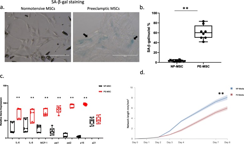
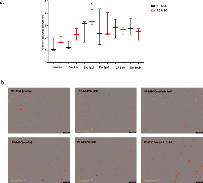
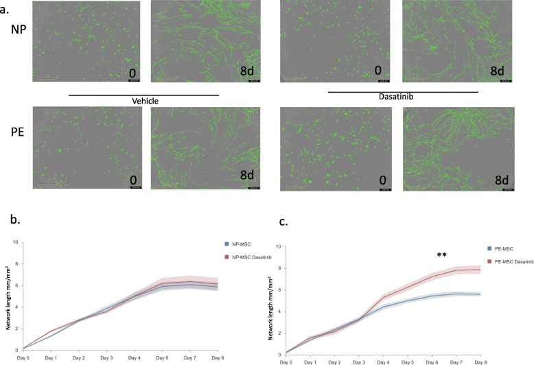
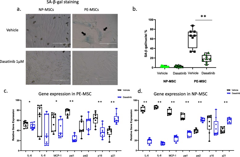
Methods: MSC were isolated from abdominal fat tissue explants removed during medically indicated C-sections from women with preeclampsia (PE-MSC, n = 10) and those with normotensive pregnancies (NP-MSC, n = 12). Sections of the frozen subcutaneous adipose tissue were assessed for inflammation by staining for tumor necrosis factor (TNF)-alpha and monocyte chemoattractant protein (MCP)-1. Viability, proliferation, and migration were compared between PE-MSC vs. NP-MSC. Apoptosis and angiogenesis were assayed before and after treatment with a senolytic agent (1 μM dasatinib) using the IncuCyte S3 Live-Cell Analysis System. Similarly, staining for senescence-associated beta galactosidase (SABG) and qPCR for gene expression of senescence markers, p16 and p21, as well as senescence-associated secretory phenotype (SASP) components, IL-6, IL-8, MCP-1, and PAI-1, were studied before and after treatment with dasatinib and compared between PE and NP.

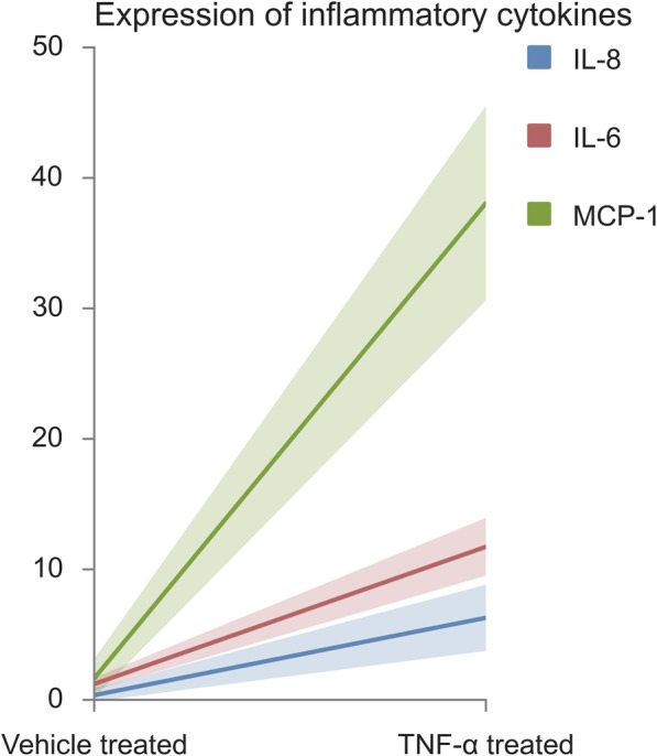
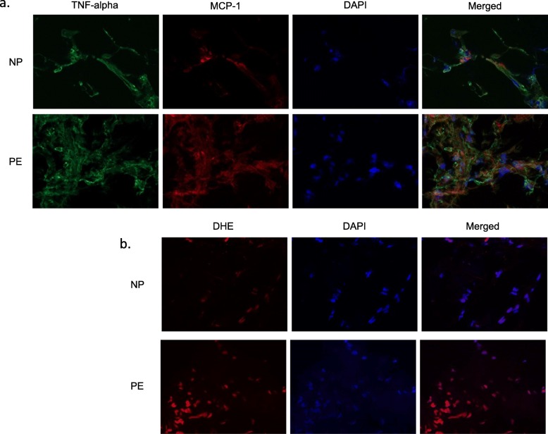
Results: After in vitro exposure to TNF-alpha, MSC demonstrated upregulation of SASP components, including interleukins-6 and -8 and MCP-1. Staining of the subcutaneous adipose tissue sections revealed a greater inflammatory response in preeclampsia, based on the higher levels of both TNF-alpha and MCP-1 compared to normotensive pregnancies (p < 0.001 and 0.024, respectively). MSC isolated from PE demonstrated a lower percentage of live MSC cells (p = 0.012), lower proliferation (p = 0.005), and higher migration (p = 0.023). At baseline, PE-MSC demonstrated a senescent phenotype, reflected by more abundant staining for SABG (p < 0.001), upregulation of senescence markers and SASP components, as well as lower angiogenic potential (p < 0.001), compared to NP-MSC. Treatment with dasatinib increased significantly the number of apoptotic PE-MSC compared to NP-MSC (0.011 vs. 0.093) and decreased the gene expression of p16 and six SASP components. The mechanistic link between senescence and impaired angiogenesis in PE was confirmed by improved angiogenic potential of PE-MSC (p < 0.001) after dasatinib treatment.

Conclusions: Our data suggest that MSC senescence exerts inhibitory effects on angiogenesis in preeclampsia. Senolytic agents may offer the opportunity for mechanism-based therapies.

Keywords: Angiogenesis, Senolytics, Dasatinib; Mesenchymal stem cells; Preeclampsia; Senescence.


Conflict of interest statement

JLK and TT have a financial interest related to this research. Patents on senolytic drugs are held by Mayo Clinic. This research has been reviewed by the Mayo Clinic Conflict of Interest Review Board and was conducted in compliance with Mayo Clinic Conflict of Interest policies. No conflicts of interest, financial or otherwise, are declared by the other authors.

Figures

Fig. 1
Fig. 1
Expression of inflammatory cytokines in non-pregnant woman MSC treated for 24 h with TNF-alpha. All three markers tested were significantly elevated after treatment with TNF-alpha vs. vehicle (presented as mean ± SD): IL-6 (in red), 11.73 ± 2.20 vs. 1.22 ± 0.47 (p = 0.009); IL-8 (in blue), 6.29 ± 2.53 vs. 0.36 ± 0.47 (p = 0.38); MCP-1 (in green), 38.07 ± 7.46 vs. 1.65 ± 1.46 (p = 0.010), respectively
Fig. 2
Fig. 2
Fat tissue staining for markers of inflammation and oxidative stress in normotensive pregnant (NP) (upper rows) and preeclamptic (PE) women (lower rows). TNF-alpha and MCP-1 were upregulated in PE. Representative images for TNF-alpha, MCP-1, and DAPI (4,6-Diamidino-2-phenylindole, dihydrochloride) nuclear (nucleus) staining, as well as merged TNF-alpha and MCP-1 (a). DHE (dihydroethidium) staining tended to be increased in PE compared to NP. Representative images for DHE and DAPI, as well as merged DHE and DAPI (b)
Fig. 3
Fig. 3
Representative flow cytometry scatterplots of MSC viability. MSC viability tested using Annexin V (channel 11) and Sytox (Channel 2) shows decreased MSC viability in preeclamptic (85%) vs. normotensive (94%) pregnancies (p = 0.01). This is a representative image where yellow panel represents live cells, red panel represents dead cells, and orange panel represents apoptotic cells
Fig. 4
Fig. 4
Senescent cell burden is higher in PE-MSC compared to NP-MSC. SABG staining revealed a higher number of stained cells (marked with black arrows) in PE-MSC compared to their normotensive counterparts (a). Data presented as mean values of SABG-stained MSC with min-max (b). Expression of p16, but not p21, was significantly increased. All SASP genes were significantly more highly expressed in PE-MSC compared to NP-MSC. Data are shown as box-plots (min-max) with all individual values (c). PE-MSC and NP-MSC were co-cultured with GFP expressing HUVEC for 8 days in total, and total network length was measured continuously every 3 h. Significantly lower angiogenic potential was registered for HUVEC co-cultured with PE-MSC, compared to NP-MSC (F = 13.965; df = 8, p < 0.001) (d)
Fig. 5
Fig. 5
Apoptotic effects of the senolytic agent, dasatinib, on MSC. Dose-response experiments showed that PE-MSCs are sensitive to a lower concentration (1 μM) of dasatinib, while the apoptotic effect of the drug is lower at higher concentrations of the drug (a). Treatment with 1 μM dasatinib revealed significant apoptotic effects in PE-MSC (p = 0.0117) compared to the non-treated cells, but not in NP-MSC (p = 0.0934). Representative image shows apoptotic cells stained red for PE-MSC and NP-MSC in all three conditions (medium, vehicle, treatment) (n = 3) (b)
Fig. 6
Fig. 6
Angiogenic potential of dasatinib-treated PE-MSC was improved after the treatment. Representative images showing the total network length formed at day 0 and day 8 after senolytic agent treatment of PE-MSC and NP-MSC (a). While no significant difference in angiogenic potential of NP-MSC (n = 9) was observed after the treatment (F = 0.406; df = 8; p = 0.916) (b), NP-MSC co-cultured HUVEC showed significant improvement in angiogenesis (F = 22.436; df = 8; p < 0.001) (c)
Fig. 7
Fig. 7
Treatment with dasatinib cleared senescent cells from PE-MSC and affected senescence-related gene expression. Representative images from SABG staining show abundant senescent cells in PE-MSC (marked with black arrows), but not in NP-MSC, in vehicle (a). Dasatinib treatment completely removed senescent cells from PE-MSC (p < 0.001) (b). PE-MSC had a significant decrease in expression of p16 (p = 0.025) and PAI-1 (p < 0.001), IL-6 (p = 0. 0487), and MCP-1 (p = 0.040), while IL-8 (p = 0.136) was modestly decreased after treatment. Significantly increased expression of the p21 and PAI-2 genes was observed after the treatment (c). Relative expression of the senescence marker gene, p16, in NP-MSC after treatment with Dasatinib remained unchanged (p = 0.136). Apart from p21 and PAI-2 whose expression increased in a manner similar to PE-MSC, the relative gene expression of the other tested genes was decreased in NP-MSC after the treatment with dasatinib (p < 0.001) (d)

Similar articles

Cited by

References

    1. Report of the National High Blood Pressure Education Program Working Group on High Blood Pressure in Pregnancy. Am J Obstet Gynecol. 2000;183(1):S1-S22. - PubMed
    1. Shohara R, Yamamoto A, Takikawa S, Iwase A, Hibi H, Kikkawa F, et al. Mesenchymal stromal cells of human umbilical cord Wharton’s jelly accelerate wound healing by paracrine mechanisms. Cytotherapy. 2012;14(10):1171–1181. doi: 10.3109/14653249.2012.706705. - DOI - PubMed
    1. Mirotsou M, Zhang ZY, Deb A, Zhang LN, Gnecchi M, Noiseux N, et al. Secreted frizzled related protein 2 (Sfrp2) is the key Akt-mesenchymal stem cell-released paracrine factor mediating myocardial survival and repair. Proc Natl Acad Sci U S A. 2007;104(5):1643–1648. doi: 10.1073/pnas.0610024104. - DOI - PMC - PubMed
    1. Gnecchi M, Zhang ZP, Ni AG, Dzau VJ. Paracrine mechanisms in adult stem cell signaling and therapy. Circ Res. 2008;103(11):1204–1219. doi: 10.1161/CIRCRESAHA.108.176826. - DOI - PMC - PubMed
    1. James JL, Srinivasan S, Alexander M, Chamley LW. Can we fix it? Evaluating the potential of placental stem cells for the treatment of pregnancy disorders. Placenta. 2014;35(2):77–84. doi: 10.1016/j.placenta.2013.12.010. - DOI - PubMed

Publication types

LinkOut - more resources