

Estimated Ventricular Size, Asthma Severity, and Exacerbations: The Severe Asthma Research Program III Cohort
- PMID: 31521672
- PMCID: PMC7005378
- DOI: 10.1016/j.chest.2019.08.2185
Estimated Ventricular Size, Asthma Severity, and Exacerbations: The Severe Asthma Research Program III Cohort
Abstract
Background: Relative enlargement of the pulmonary artery (PA) on chest CT imaging is associated with respiratory exacerbations in patients with COPD or cystic fibrosis. We sought to determine whether similar findings were present in patients with asthma and whether these findings were explained by differences in ventricular size.
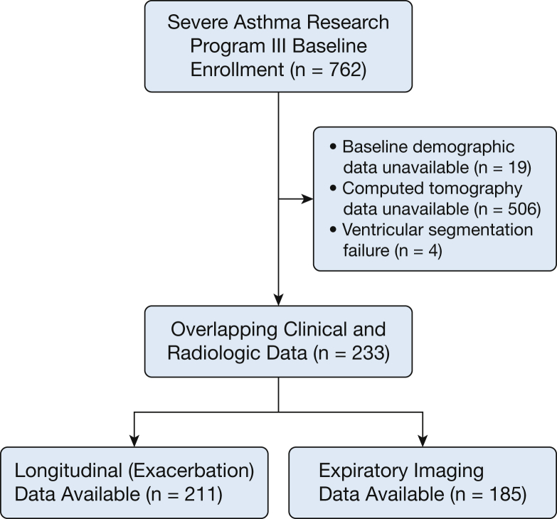
Methods: We measured the PA and aorta diameters in 233 individuals from the Severe Asthma Research Program III cohort. We also estimated right, left, and total epicardial cardiac ventricular volume indices (eERVVI, eELVVI, and eETVVI, respectively). Associations between the cardiac and PA measures (PA-to-aorta [PA/A] ratio, eERVVI-to-eELVVI [eRV/eLV] ratio, eERVVI, eELVVI, eETVVI) and clinical measures of asthma severity were assessed by Pearson correlation, and associations with asthma severity and exacerbation rate were evaluated by multivariable linear and zero-inflated negative binomial regression.
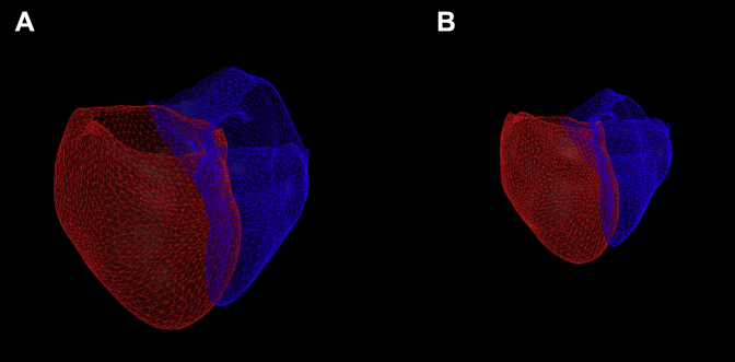
Results: Asthma severity was associated with smaller ventricular volumes. For example, those with severe asthma had 36.1 mL/m2 smaller eETVVI than healthy control subjects (P = .003) and 14.1 mL/m2 smaller eETVVI than those with mild/moderate disease (P = .011). Smaller ventricular volumes were also associated with a higher rate of asthma exacerbations, both retrospectively and prospectively. For example, those with an eETVVI less than the median had a 57% higher rate of exacerbations during follow-up than those with eETVVI greater than the median (P = .020). Neither PA/A nor eRV/eLV was associated with asthma severity or exacerbations.
Conclusions: In patients with asthma, smaller cardiac ventricular size may be associated with more severe disease and a higher rate of asthma exacerbations.
Trial registry: ClinicalTrials.gov; No.: NCT01761630; URL: www.clinicaltrials.gov.
Keywords: CT imaging; asthma; heart.
Copyright © 2019 American College of Chest Physicians. Published by Elsevier Inc. All rights reserved.
Figures
Comment in
-
Estimated Ventricular Size: A New Predictor of Asthma Severity and Exacerbation Rate?Chest. 2020 Feb;157(2):243-244. doi: 10.1016/j.chest.2019.10.019. Chest. 2020. PMID: 32033641 No abstract available.
References
Publication types
MeSH terms
Associated data
Grants and funding
- R01 HL122464/HL/NHLBI NIH HHS/United States
- UL1 TR000427/TR/NCATS NIH HHS/United States
- K23 HL136905/HL/NHLBI NIH HHS/United States
- U01 HL146002/HL/NHLBI NIH HHS/United States
- R01 HL116473/HL/NHLBI NIH HHS/United States
- U10 HL109250/HL/NHLBI NIH HHS/United States
- L30 HL134158/HL/NHLBI NIH HHS/United States
- R01 HL122531/HL/NHLBI NIH HHS/United States
- UL1 TR001420/TR/NCATS NIH HHS/United States
- U10 HL109172/HL/NHLBI NIH HHS/United States
- U10 HL109257/HL/NHLBI NIH HHS/United States
- K23 HL114735/HL/NHLBI NIH HHS/United States
- T32 HL007633/HL/NHLBI NIH HHS/United States
- S10 OD018526/OD/NIH HHS/United States
- R01 HL116931/HL/NHLBI NIH HHS/United States
- K23 AI125785/AI/NIAID NIH HHS/United States
- U10 HL109164/HL/NHLBI NIH HHS/United States
- UL1 TR000448/TR/NCATS NIH HHS/United States
- UL1 TR002373/TR/NCATS NIH HHS/United States
- P30 DK054759/DK/NIDDK NIH HHS/United States
- U10 HL109086/HL/NHLBI NIH HHS/United States
- U10 HL109168/HL/NHLBI NIH HHS/United States
- K08 HL145118/HL/NHLBI NIH HHS/United States
- UL1 TR001102/TR/NCATS NIH HHS/United States
- UL1 TR002345/TR/NCATS NIH HHS/United States
- U10 HL109152/HL/NHLBI NIH HHS/United States
- U10 HL109146/HL/NHLBI NIH HHS/United States
