

doi: 10.1016/j.ipej.2019.09.003.
Epub 2019 Sep 12.
A curious case of impossible coronary sinus cannulation
Affiliations
- PMID: 31521673
- PMCID: PMC6823679
- DOI: 10.1016/j.ipej.2019.09.003
Item in Clipboard
A curious case of impossible coronary sinus cannulation
Indian Pacing Electrophysiol J.
2019 Sep-Oct.
Abstract
A 40-year-old male, diagnosed to have WPW syndrome and symptomatic with recurrent palpitations, was taken up for radiofrequency ablation. There was difficulty in coronary sinus cannulation. Coronary venogram revealed coronary sinus atresia with persistent left superior vena cava, and collateral venous pathways draining into the right atrium. This case is discussed for the rare coronary venous anomaly, its embryology and the difficulties in the management during electrophysiological studies.
Copyright © 2019 Indian Heart Rhythm Society. Production and hosting by Elsevier B.V. All rights reserved.
Figures
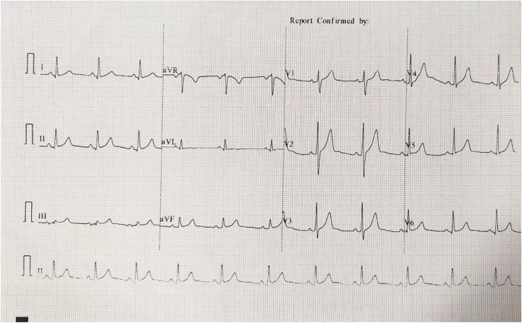
Baseline 12-lead ECG of patient – negative delta wave in Lead I, no transition of delta wave suggestive of a left-lateral pathway.
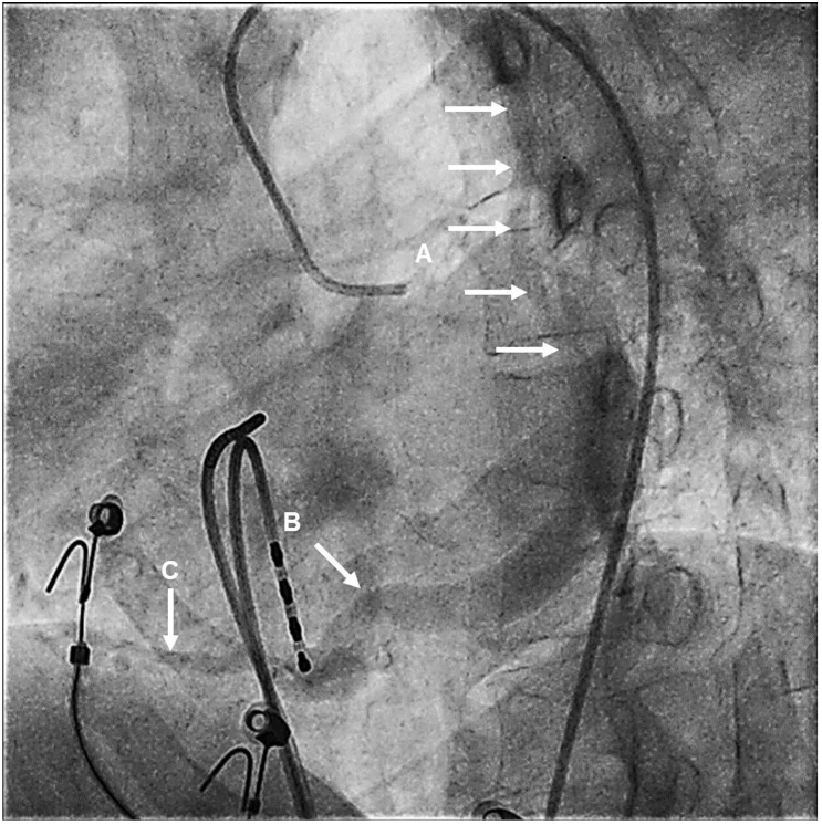
Venous phase of the left coronary angiogram in left anterior oblique view – (A) Persistent left superior vena cava (multiple arrows) (B) Absence of coronary sinus drainage into the right atrium (C) Collateral pathways from coronary sinus to the right atrium.
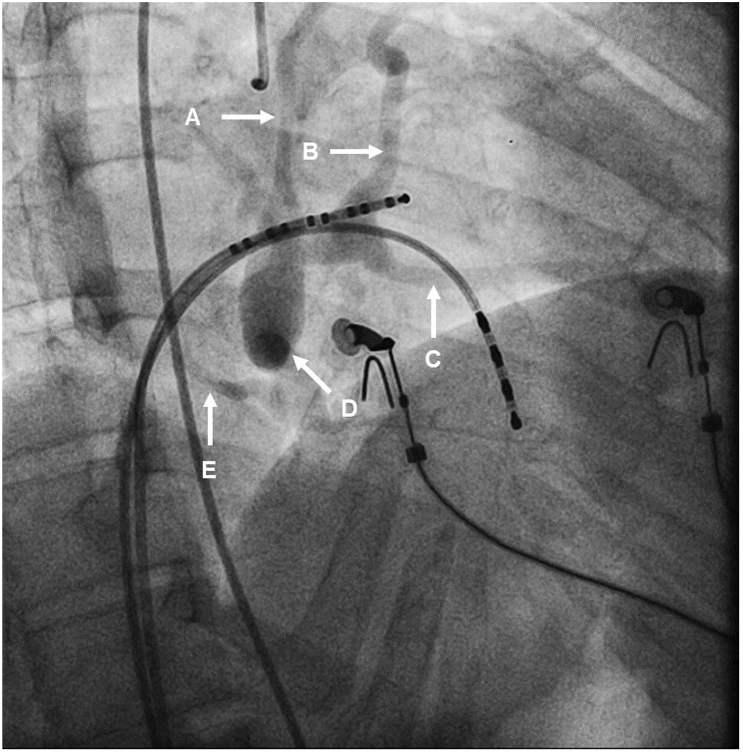
Venous phase of the left coronary angiogram in right anterior oblique view – (A) Persistent left superior vena cava (B) Coronary sinus (C) Marginal vein (D) Absence of coronary sinus drainage into the right atrium (E) Collateral pathways from coronary sinus to the right atrium.
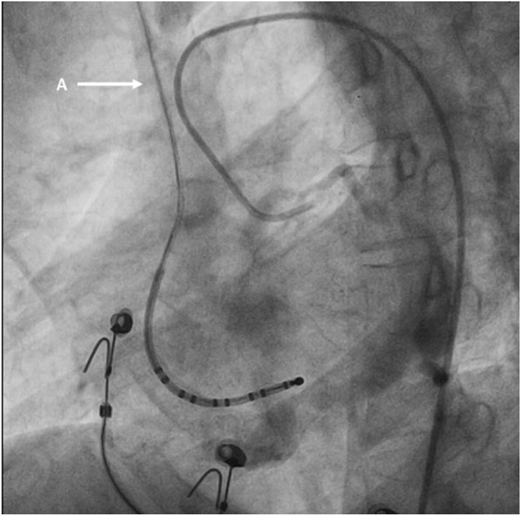
Venous phase of the left coronary angiogram in left anterior oblique view – (A) Right superior vena cava, filling from left superior vena cava through connecting cava (not seen in the picture) and draining into the right atrium.
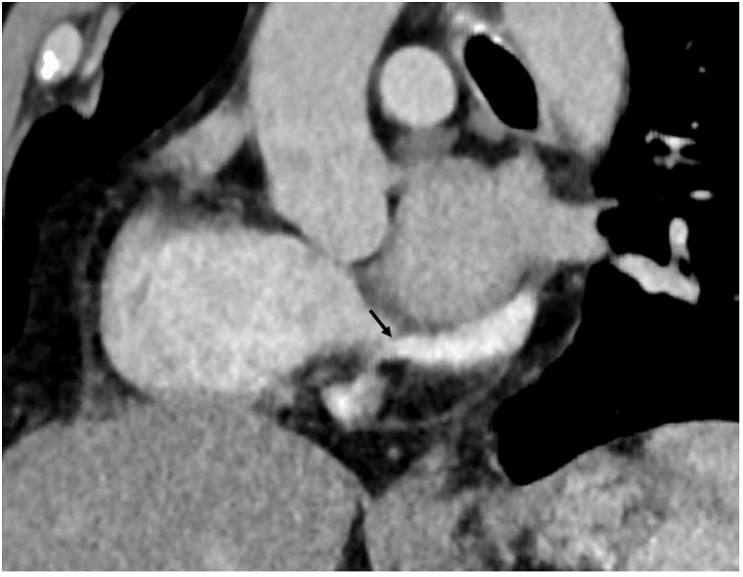
Computed tomography – coronal section – Black arrow pointing to the membrane at the coronary sinus. Note the higher opacification in the coronary sinus compared to the right atrium.
References
-
- Shum J.S., Kim S.M., Choe Y.H. Multidetector CT and MRI of ostial atresia of the coronary sinus, associated collateral venous pathways and cardiac anomalies. Clin Radiol. 2012;67(12):e47–e52. - PubMed
-
- Santoscoy R., Walters H.L., 3rd, Ross R.D., Lyons J.M., Hakimi M. Coronary sinus ostial atresia with persistent left superior vena cava. Ann Thorac Surg. 1996;61(3):879–882. - PubMed
-
- Saremi F., Muresian H., Sánchez-Quintana D. Coronary veins: comprehensive CT-anatomic classification and review of variants and clinical implications. RadioGraphics. 2012;32(1):E1–E32. - PubMed
-
- Paul J.J., Williams R.V., Minich L.L.A., Tani L.Y. Echocardiographic diagnosis of coronary sinus ostial atresia. J Am Soc Echocardiogr. 2002;15(9):991–993. - PubMed
-
- Qanadli S.D., Rolf T., Glauser F., Delay D., Beigelman-Aubry C., Pretre R. Coronary sinus atresia with persistent left superior vena cava: unusual clinical presentation and endovascular management. Cardiovasc Interv Radiol. 2014;37(3):825–828. - PubMed
LinkOut - more resources
Full Text Sources