. 2019 Dec:203:116192.
doi: 10.1016/j.neuroimage.2019.116192. Epub 2019 Sep 12.

Imaging the human hippocampus with optically-pumped magnetoencephalography

Affiliations

Imaging the human hippocampus with optically-pumped magnetoencephalography

Daniel N Barry et al. Neuroimage. 2019 Dec.

Abstract

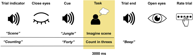
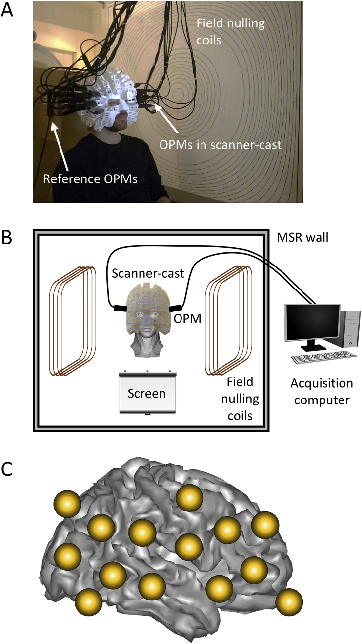
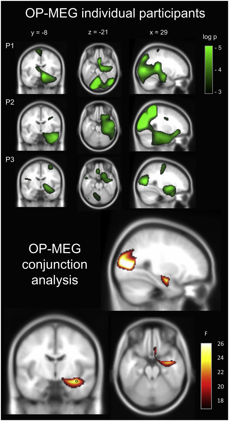
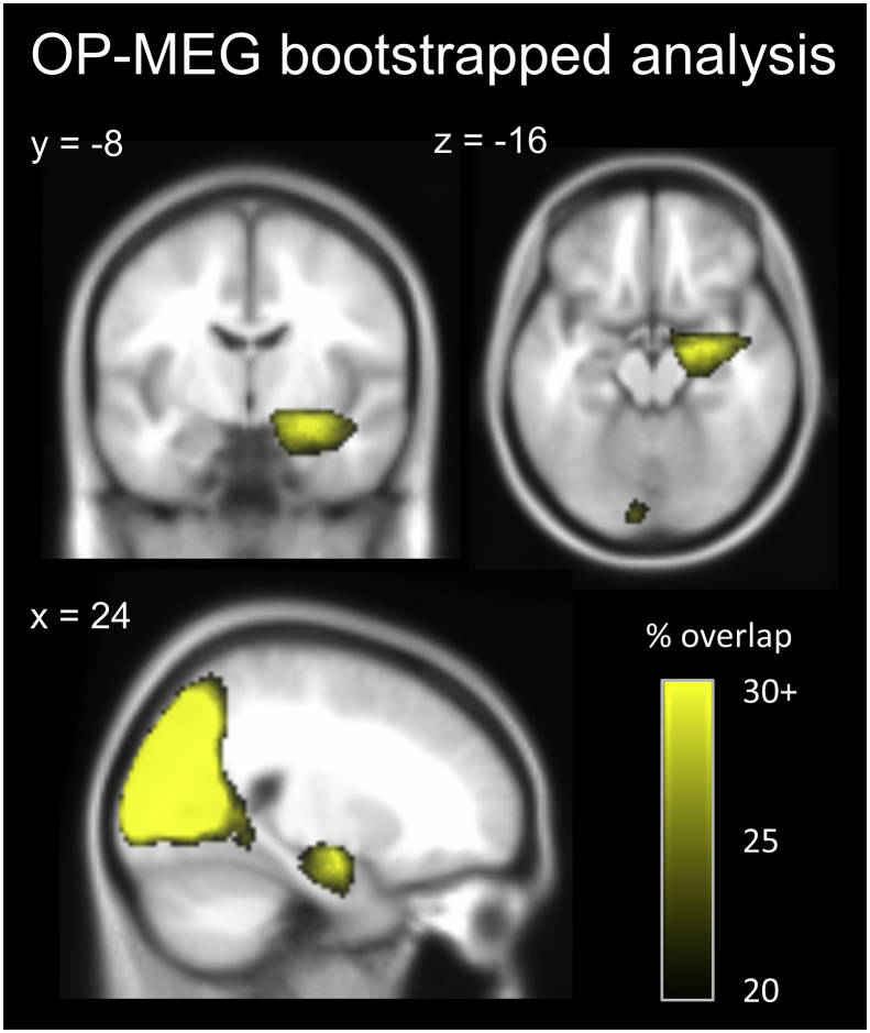
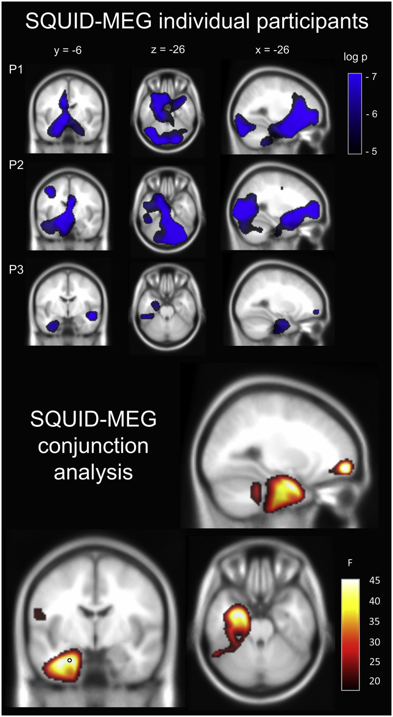
Optically-pumped (OP) magnetometers allow magnetoencephalography (MEG) to be performed while a participant's head is unconstrained. To fully leverage this new technology, and in particular its capacity for mobility, the activity of deep brain structures which facilitate explorative behaviours such as navigation, must be detectable using OP-MEG. One such crucial brain region is the hippocampus. Here we had three healthy adult participants perform a hippocampal-dependent task - the imagination of novel scene imagery - while being scanned using OP-MEG. A conjunction analysis across these three participants revealed a significant change in theta power in the medial temporal lobe. The peak of this activated cluster was located in the anterior hippocampus. We repeated the experiment with the same participants in a conventional SQUID-MEG scanner and found similar engagement of the medial temporal lobe, also with a peak in the anterior hippocampus. These OP-MEG findings indicate exciting new opportunities for investigating the neural correlates of a range of crucial cognitive functions in naturalistic contexts including spatial navigation, episodic memory and social interactions.

Keywords: Hippocampus; Imagination; Magnetoencephalography; Optically-pumped magnetometers; Scene construction; Source localisation.


Figures

Fig. 1
Fig. 1
Trial structure. The task period used in the analyses is highlighted.
Fig. 2
Fig. 2
The OP-MEG setup for studying hippocampal engagement during the construction of scene imagery. (A) The participant was seated inside a magnetically shielded room wearing the scanner-cast with OP-MEG sensors inserted into slots. (B) The field nulling coils were placed either side of the participant and conferred motion robustness to the recordings by nulling the field over a 40 × 40 × 40 cm3 volume within which head movement was tolerated. (C) The OP-MEG array covered inferior and superior occipital, temporal and frontal areas bilaterally.
Fig. 3
Fig. 3
Results of the OP-MEG study. Top panel: Source level theta (4–8 Hz) power changes for each participant during the imagination of novel scenes. P = participant. Images are superimposed on the MNI 152 T1 image. Images are thresholded at a significance level of p < 0.05 (uncorrected for display purposes). Bottom panel: A conjunction analysis of the three participants revealed significant activation of the right anterior hippocampus. The location of peak activity in the hippocampus is indicated with a black circle. Task-based modulation of theta power was also observed in the occipital lobe, with the peak of this large cluster localised to the cuneus. This is not surprising, given the comparison of rich scene imagery with the low level counting baseline task which had no imagery requirement. Previous neuroimaging studies have documented similar engagement of visual cortex during imagery as that evoked by perception of the same content (Albers et al., 2013; Nishimura et al., 2015). Images are FDR thresholded at q < 0.005. This result can also be examined here: https://doi.org/10.6084/m9.figshare.8681483.
Fig. 4
Fig. 4
Results of the bootstrapped OP-MEG conjunction analyses. The conjunction analysis was repeated 500 times with sub-sampling of each participant’s trials to give an indication of the stability of the observed significant clusters. Within the medial temporal lobe, the location of the highest concentration of conjunctions where voxels displayed significant activity was in the right anterior hippocampus. Occipital regions which emerged from the original conjunction analysis were also reliably engaged. The map of the percentage of significant conjunctions is superimposed on the MNI 152 T1 image, at an arbitrarily chosen minimum threshold of 20% overlap across all conjunctions.
Fig. 5
Fig. 5
Results of the SQUID-MEG study. Top panel: Source level theta (4–8 Hz) power changes for each participant during the imagination of novel scenes. P = participant. Images are superimposed on the MNI 152 T1 image. Images are thresholded at a significance level of p < 0.007 (uncorrected for display purposes). Bottom panel: A conjunction analysis of the three participants revealed significant activation of the left medial temporal lobe. The peak of this cluster was located in the anterior hippocampus (indicated with a black circle). Task-based modulation of theta power was also observed in the left frontal gyrus. Images are FDR thresholded at q < 0.005. This result can also be examined here: https://doi.org/10.6084/m9.figshare.8681483.

References

    1. Addis D.R., Wong A.T., Schacter D.L. Remembering the past and imagining the future: common and distinct neural substrates during event construction and elaboration. Neuropsychologia. 2007;45(7):1363–1377. doi: 10.1016/j.neuropsychologia.2006.10.016. - DOI - PMC - PubMed
    1. Addis D.R., Cheng T., Roberts R.P., Schacter D.L. Hippocampal contributions to the episodic simulation of specific and general future events. Hippocampus. 2011;21(10):1045–1052. doi: 10.1002/hipo.20870. - DOI - PMC - PubMed
    1. Albers A.M., Kok P., Toni I., Dijkerman H.C., de Lange F.P. Shared representations for working memory and mental imagery in early visual cortex. Curr. Biol. 2013;23:1427–1431. doi: 10.1016/j.cub.2013.05.065. - DOI - PubMed
    1. Attal Y., Schwartz D. Assessment of subcortical source localization using deep brain activity imaging model with minimum norm operators: a meg study. PLoS One. 2013;8(3) doi: 10.1371/journal.pone.0059856. - DOI - PMC - PubMed
    1. Attal Y., Maess B., Friederici A., David O. Head models and dynamic causal modeling of subcortical activity using magnetoencephalographic/electroencephalographic data. Rev. Neurosci. 2012;23(1):85–95. doi: 10.1515/rns.2011.056. - DOI - PubMed

Publication types