

Probing RNA structure in vivo
- PMID: 31521910
- PMCID: PMC6888943
- DOI: 10.1016/j.sbi.2019.07.008
Probing RNA structure in vivo
Abstract
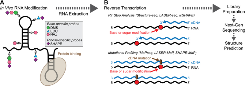
RNA structure underpins many essential functions in biology. New chemical reagents and techniques for probing RNA structure in living cells have emerged in recent years. High-throughput, genome-wide techniques such as Structure-seq2 and DMS-MaPseq exploit nucleobase modification by dimethylsulfate (DMS) to obtain complete structuromes, and are applicable to multiple domains of life and conditions. New reagents such as 1-ethyl-3-(3-dimethylaminopropyl)carbodiimide (EDC), glyoxal, and nicotinoyl azide (NAz) greatly expand the capabilities of nucleobase probing in cells. Additionally, ribose-targeting reagents in selective 2'-hydroxyl acylation and primer extension (SHAPE) detect RNA flexibility in vivo. These techniques, coupled with crosslinking nucleobases in psoralen analysis of RNA interactions and structures (PARIS), provide new and diverse ways to elucidate RNA secondary and tertiary structure in vivo and genome-wide.
Copyright © 2019 Elsevier Ltd. All rights reserved.
Figures


References
-
- Noller HF, Hoffarth V, Zimniak L: Unusual resistance of peptidyl transferase to protein extraction procedures. Science 1992, 256:1416–1419. - PubMed
-
- Yusupov MM, Yusupova GZ, Baucom A, Lieberman K, Earnest TN, Cate JH, Noller HF: Crystal structure of the ribosome at 5.5 A resolution. Science 2001, 292:883–896. - PubMed
-
- Yanofsky C: Attenuation in the control of expression of bacterial operons. Nature 1981, 289:751–758. - PubMed
-
- Altuvia S, Kornitzer D, Teff D, Oppenheim AB: Alternative mRNA structures of the cIII gene of bacteriophage lambda determine the rate of its translation initiation. J Mol Biol 1989, 210:265–280. - PubMed
-
- Naville M, Gautheret D: Transcription attenuation in bacteria: theme and variations. Brief Funct Genomics 2010, 9:178–189. - PubMed
Publication types
MeSH terms
Substances
Grants and funding
LinkOut - more resources
Full Text Sources
