

Automated Echocardiographic Quantification of Left Ventricular Ejection Fraction Without Volume Measurements Using a Machine Learning Algorithm Mimicking a Human Expert
- PMID: 31522550
- PMCID: PMC7099856
- DOI: 10.1161/CIRCIMAGING.119.009303
Automated Echocardiographic Quantification of Left Ventricular Ejection Fraction Without Volume Measurements Using a Machine Learning Algorithm Mimicking a Human Expert
Abstract
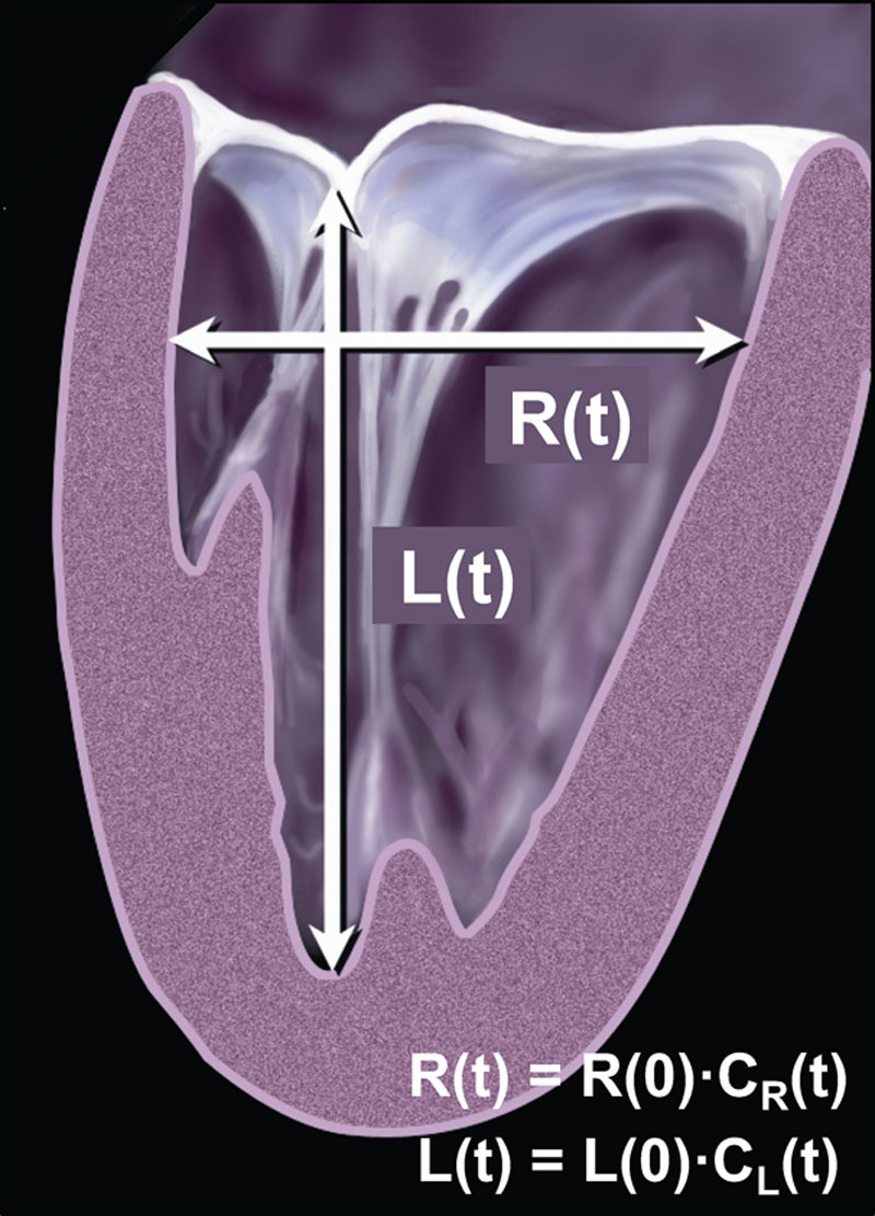
Background: Echocardiographic quantification of left ventricular (LV) ejection fraction (EF) relies on either manual or automated identification of endocardial boundaries followed by model-based calculation of end-systolic and end-diastolic LV volumes. Recent developments in artificial intelligence resulted in computer algorithms that allow near automated detection of endocardial boundaries and measurement of LV volumes and function. However, boundary identification is still prone to errors limiting accuracy in certain patients. We hypothesized that a fully automated machine learning algorithm could circumvent border detection and instead would estimate the degree of ventricular contraction, similar to a human expert trained on tens of thousands of images.
Methods: Machine learning algorithm was developed and trained to automatically estimate LVEF on a database of >50 000 echocardiographic studies, including multiple apical 2- and 4-chamber views (AutoEF, BayLabs). Testing was performed on an independent group of 99 patients, whose automated EF values were compared with reference values obtained by averaging measurements by 3 experts using conventional volume-based technique. Inter-technique agreement was assessed using linear regression and Bland-Altman analysis. Consistency was assessed by mean absolute deviation among automated estimates from different combinations of apical views. Finally, sensitivity and specificity of detecting of EF ≤35% were calculated. These metrics were compared side-by-side against the same reference standard to those obtained from conventional EF measurements by clinical readers.
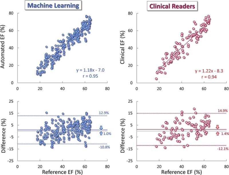
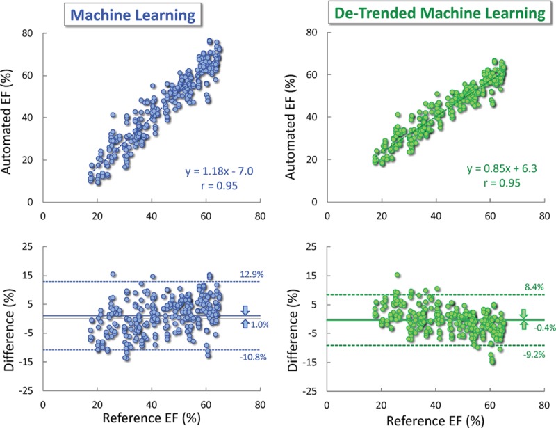
Results: Automated estimation of LVEF was feasible in all 99 patients. AutoEF values showed high consistency (mean absolute deviation =2.9%) and excellent agreement with the reference values: r=0.95, bias=1.0%, limits of agreement =±11.8%, with sensitivity 0.90 and specificity 0.92 for detection of EF ≤35%. This was similar to clinicians' measurements: r=0.94, bias=1.4%, limits of agreement =±13.4%, sensitivity 0.93, specificity 0.87.
Conclusions: Machine learning algorithm for volume-independent LVEF estimation is highly feasible and similar in accuracy to conventional volume-based measurements, when compared with reference values provided by an expert panel.
Keywords: echocardiography; endocardium; left ventricular function; machine learning; observer variation.
Figures
Comment in
-
Combining Artificial Intelligence With Human Insight to Automate Echocardiography.Circ Cardiovasc Imaging. 2019 Sep;12(9):e009727. doi: 10.1161/CIRCIMAGING.119.009727. Epub 2019 Sep 16. Circ Cardiovasc Imaging. 2019. PMID: 31522554 Free PMC article. No abstract available.
References
-
- Lang RM, Badano LP, Mor-Avi V, Afilalo J, Armstrong A, Ernande L, Flachskampf FA, Foster E, Goldstein SA, Kuznetsova T, Lancellotti P, Muraru D, Picard MH, Rietzschel ER, Rudski L, Spencer KT, Tsang W, Voigt JU. Recommendations for cardiac chamber quantification by echocardiography in adults: an update from the American society of echocardiography and the European Association of cardiovascular imaging. J Am Soc Echocardiogr. 2015;28:1.e14–39.e14. doi: 10.1016/j.echo.2014.10.003 - PubMed
-
- Mor-Avi V, Jenkins C, Kühl HP, Nesser HJ, Marwick T, Franke A, Ebner C, Freed BH, Steringer-Mascherbauer R, Pollard H, Weinert L, Niel J, Sugeng L, Lang RM. Real-time 3-dimensional echocardiographic quantification of left ventricular volumes: multicenter study for validation with magnetic resonance imaging and investigation of sources of error. JACC Cardiovasc Imaging. 2008;1:413–423. doi: 10.1016/j.jcmg.2008.02.009 - PubMed
-
- Rich S, Sheikh A, Gallastegui J, Kondos GT, Mason T, Lam W. Determination of left ventricular ejection fraction by visual estimation during real-time two-dimensional echocardiography. Am Heart J. 1982;104:603–606. doi: 10.1016/0002-8703(82)90233-2 - PubMed
-
- Hope MD, de la Pena E, Yang PC, Liang DH, McConnell MV, Rosenthal DN. A visual approach for the accurate determination of echocardiographic left ventricular ejection fraction by medical students. J Am Soc Echocardiogr. 2003;16:824–831. doi: 10.1067/S0894-7317(03)00400-0 - PubMed
-
- Mele D, Campana M, Sclavo M, Seveso G, Aschieri D, Nesta F, D’Aiello I, Ferrari R, Levine RA. Impact of tissue harmonic imaging in patients with distorted left ventricles: improvement in accuracy and reproducibility of visual, manual and automated echocardiographic assessment of left ventricular ejection fraction. Eur J Echocardiogr. 2003;4:59–67. doi: 10.1053/euje.2002.0619 - PubMed
Publication types
MeSH terms
LinkOut - more resources
Full Text Sources
Other Literature Sources
