. 2019:105:93-116.
doi: 10.1016/bs.aivir.2019.08.002. Epub 2019 Aug 22.

Structural insights into coronavirus entry

Affiliations

Structural insights into coronavirus entry

M Alejandra Tortorici et al. Adv Virus Res. 2019.

Abstract

Coronaviruses (CoVs) have caused outbreaks of deadly pneumonia in humans since the beginning of the 21st century. The severe acute respiratory syndrome coronavirus (SARS-CoV) emerged in 2002 and was responsible for an epidemic that spread to five continents with a fatality rate of 10% before being contained in 2003 (with additional cases reported in 2004). The Middle-East respiratory syndrome coronavirus (MERS-CoV) emerged in the Arabian Peninsula in 2012 and has caused recurrent outbreaks in humans with a fatality rate of 35%. SARS-CoV and MERS-CoV are zoonotic viruses that crossed the species barrier using bats/palm civets and dromedary camels, respectively. No specific treatments or vaccines have been approved against any of the six human coronaviruses, highlighting the need to investigate the principles governing viral entry and cross-species transmission as well as to prepare for zoonotic outbreaks which are likely to occur due to the large reservoir of CoVs found in mammals and birds. Here, we review our understanding of the infection mechanism used by coronaviruses derived from recent structural and biochemical studies.

Keywords: Coronavirus; Fusion protein; Membrane fusion; Proteolytic activation; Spike glycoprotein; Vaccine design.


Figures

Fig. 1
Fig. 1
CryoEM structure of the apo-HCoV-OC43 S glycoprotein. (A) Ribbon diagrams of the apo HCoV-OC43 S ectodomain trimer (PDB: 6OHW) in two orthogonal orientations, from the side (left) and from the top, looking towards the viral membrane (right). (B) Side view of one S protomer. (C) Ribbon diagram of the HCoV-OC43 S1 subunit. (D–E) Close-up view of HCoV-OC43 domain A (D) and domain B (E). (F) Ribbon diagram of the HCoV-OC43 S2 subunit in the prefusion conformation. The N- and C-termini are labeled in panels (B–E).
Fig. 2
Fig. 2
Structural studies of human CoV attachment to host receptors. (A–E), Ribbon diagrams of the complex between domain A of HCoV-OC43 S with a 9-O-Ac-Sia receptor analogue ((A) PDB: 6NZK), or the domain B of SARS-CoV S with ACE2 ((B) PDB: 2AJF), HCoV-NL63 S with ACE2 ((C) PDB: 3KBH), MERS-CoV S with DPP4 ((D) PDB: 4L72) and HCoV-229E S with APN ((E) PDB: 6ATK). In panels (B–E), each domain B is rendered in light blue and the receptor binding-motifs are colored purple.
Fig. 3
Fig. 3
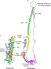
CoV S conformational changes driving the fusion reaction. (A), Ribbon diagram of the MHV S2 subunit in the prefusion conformation, PDB: 3JCL. (B), Ribbon diagram of the MHV S2 subunit in the postfusion conformation, PDB: 6B3O. The prefusion to postfusion transition involves a “jack-knife” refolding of the HR1 helices and intervening regions into a single continuous helix appended to the central helix. The connector domain and HR2 in the prefusion structure and the fusion peptide in the postfusion structure of MHV were not resolved and are therefore not shown.
Fig. 4
Fig. 4
CryoEM structures of the SARS-CoV S glycoprotein in complex with the S230 neutralizing antibody. (A–B), Molecular surface representation of a complex with one open, one partially open, and one closed B domain, PDB: 6NB6 (left) and with three open B domains that do not follow threefold symmetry, PDB: 6NB7 (right). The structures are rendered with different colors for each S protomer (light blue, plum and gold) and the S230 Fab heavy (dark magenta) and light (magenta) chains (only the variable domains are shown).
Fig. 5
Fig. 5
Organization of the HCoV-NL63 S glycan shield. Ribbon representation of the S ectodomain trimer with N-linked glycans rendered as dark-blue spheres, PDB: 5SZS.

Similar articles

Cited by

References

    1. Bai X.C., Fernandez I.S., McMullan G., Scheres S.H. Ribosome structures to near-atomic resolution from thirty thousand cryo-EM particles. elife. 2013;2 - PMC - PubMed
    1. Bakkers M.J., Lang Y., Feitsma L.J., Hulswit R.J., de Poot S.A., van Vliet A.L., Margine I., de Groot-Mijnes J.D., van Kuppeveld F.J., Langereis M.A., Huizinga E.G., de Groot R.J. Betacoronavirus adaptation to humans involved progressive loss of hemagglutinin-esterase lectin activity. Cell Host Microbe. 2017;21:356. - PMC - PubMed
    1. Bakkers M.J., Zeng Q., Feitsma L.J., Hulswit R.J., Li Z., Westerbeke A., van Kuppeveld F.J., Boons G.J., Langereis M.A., Huizinga E.G., de Groot R.J. Coronavirus receptor switch explained from the stereochemistry of protein-carbohydrate interactions and a single mutation. Proc. Natl. Acad. Sci. U. S. A. 2016;113 - PMC - PubMed
    1. Barlan A., Zhao J., Sarkar M.K., Li K., McCray P.B., Perlman S., Gallagher T. Receptor variation and susceptibility to Middle East respiratory syndrome coronavirus infection. J. Virol. 2014;88:4953. - PMC - PubMed
    1. Belouzard S., Chu V.C., Whittaker G.R. Activation of the SARS coronavirus spike protein via sequential proteolytic cleavage at two distinct sites. Proc. Natl. Acad. Sci. U. S. A. 2009;106:5871. - PMC - PubMed