

A Correlation between Wnt/Beta-catenin Signaling and the Rate of Dentin Secretion
- PMID: 31522810
- PMCID: PMC10900857
- DOI: 10.1016/j.joen.2019.07.014
A Correlation between Wnt/Beta-catenin Signaling and the Rate of Dentin Secretion
Abstract
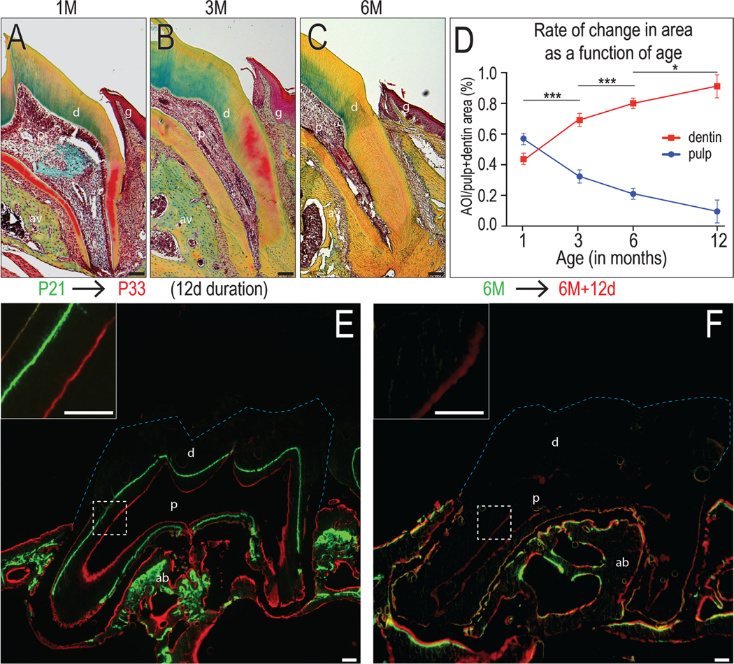
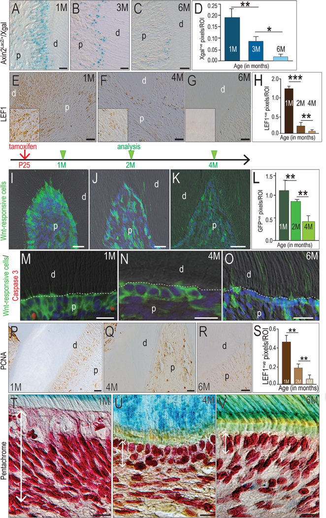
Introduction: Odontoblasts produce dentin throughout life and in response to trauma. The purpose of this study was to identify the roles of endogenous Wnt signaling in regulating the rate of dentin accumulation.
Methods: Histology, immunohistochemistry, vital dye labeling, and histomorphometric assays were used to quantify the rate of dentin accumulation as a function of age. Two strains of Wnt reporter mice were used to identify and follow the distribution and number of Wnt-responsive odontoblasts as a function of age. To show a causal relationship between dentin secretion and Wnt signaling, dentin accumulation was monitored in a strain of mice in which Wnt signaling was aberrantly elevated.
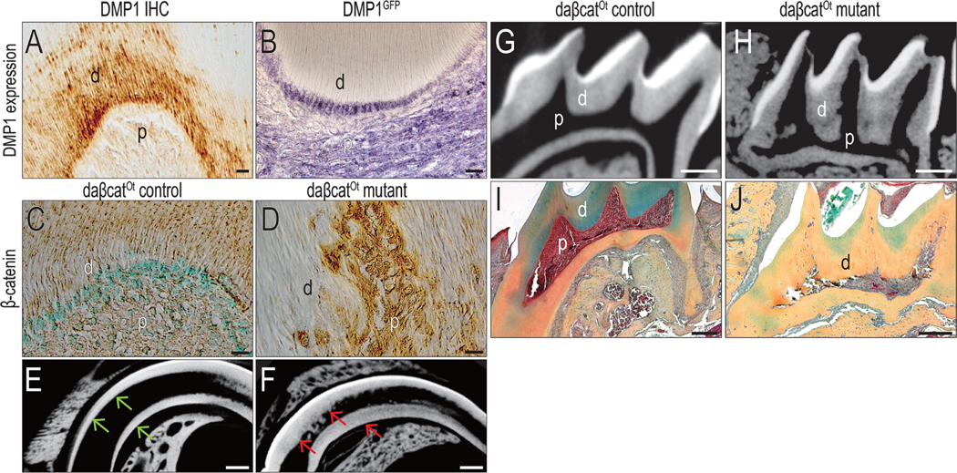
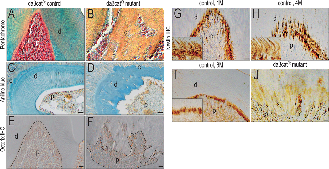
Results: Dentin deposition occurs throughout life, but the rate of accumulation slows with age. This decline in dentin secretion correlates with a decrease in endogenous Wnt signaling. In a genetically modified strain of mice, instead of tubular dentin, aberrantly elevated Wnt signaling resulted in accumulation of reparative dentin or osteodentin secreted from predontoblasts.
Conclusions: Wnt signaling regulates dentin secretion by odontoblasts, and the formation of reparative or osteodentin is the direct consequence of elevated Wnt signaling. These preclinical data have therapeutic implications for the development of a biologically based pulp capping medicant.
Keywords: Aging; Wnt signal; odontoblasts; pulp.
Copyright © 2019 American Association of Endodontists. Published by Elsevier Inc. All rights reserved.
Figures
Similar articles
-
Wnt-Responsive Odontoblasts Secrete New Dentin after Superficial Tooth Injury.J Dent Res. 2018 Aug;97(9):1047-1054. doi: 10.1177/0022034518763151. Epub 2018 Mar 22. J Dent Res. 2018. PMID: 29566345 Free PMC article.
-
The role of canonical Wnt signaling in dentin bridge formation.J Oral Biosci. 2021 Jun;63(2):199-209. doi: 10.1016/j.job.2021.03.003. Epub 2021 Apr 15. J Oral Biosci. 2021. PMID: 33845204
-
Constitutive stabilization of ß-catenin in the dental mesenchyme leads to excessive dentin and cementum formation.Biochem Biophys Res Commun. 2011 Sep 9;412(4):549-55. doi: 10.1016/j.bbrc.2011.07.116. Epub 2011 Aug 10. Biochem Biophys Res Commun. 2011. PMID: 21854758
-
Modulators of Wnt Signaling Pathway Implied in Dentin Pulp Complex Engineering: A Literature Review.Int J Mol Sci. 2022 Sep 13;23(18):10582. doi: 10.3390/ijms231810582. Int J Mol Sci. 2022. PMID: 36142496 Free PMC article. Review.
-
Odontoblasts: Specialized hard-tissue-forming cells in the dentin-pulp complex.Congenit Anom (Kyoto). 2016 Jul;56(4):144-53. doi: 10.1111/cga.12169. Congenit Anom (Kyoto). 2016. PMID: 27131345 Review.
Cited by
-
Wnt/β-Catenin Signaling Promotes the Formation of Preodontoblasts In Vitro.J Dent Res. 2021 Apr;100(4):387-396. doi: 10.1177/0022034520967353. Epub 2020 Oct 26. J Dent Res. 2021. PMID: 33103548 Free PMC article.
-
Physiologic dentin regeneration: its past, present, and future perspectives.Front Physiol. 2023 Dec 11;14:1313927. doi: 10.3389/fphys.2023.1313927. eCollection 2023. Front Physiol. 2023. PMID: 38148896 Free PMC article. Review.
-
Wnt/β-catenin Signaling Controls Maxillofacial Hyperostosis.J Dent Res. 2022 Jul;101(7):793-801. doi: 10.1177/00220345211067705. Epub 2022 Feb 3. J Dent Res. 2022. PMID: 35114849 Free PMC article.
-
Higher immunoexpression of CK14 from the Wnt-1/β-catenin pathway in the development of odontomas.Braz Dent J. 2023 Nov-Dec;34(6):110-120. doi: 10.1590/0103-6440202305452. Braz Dent J. 2023. PMID: 38133085 Free PMC article.
-
Pulp response following direct pulp capping with Tideglusib and mineral trioxide aggregate: an animal study.BMC Oral Health. 2025 Jul 19;25(1):1218. doi: 10.1186/s12903-025-06546-6. BMC Oral Health. 2025. PMID: 40684155 Free PMC article.
References
-
- Smith AJ, Cassidy N, Perry H, Begue-Kirn C, Ruch JV, Lesot H. Reactionary dentinogenesis. Int J Dev Biol 1995;39(1):273–280. - PubMed
-
- Tomes CR. On the Structure and Development of Vascular Dentine. Philosophical Transactions of the Royal Society of London 1878;169:25–47.
