. 2019 Oct 8;13(4):627-641.
doi: 10.1016/j.stemcr.2019.08.007. Epub 2019 Sep 12.

Robust, Long-Term Culture of Endoderm-Derived Hepatic Organoids for Disease Modeling

Affiliations

Robust, Long-Term Culture of Endoderm-Derived Hepatic Organoids for Disease Modeling

Soheil Akbari et al. Stem Cell Reports. .

Abstract

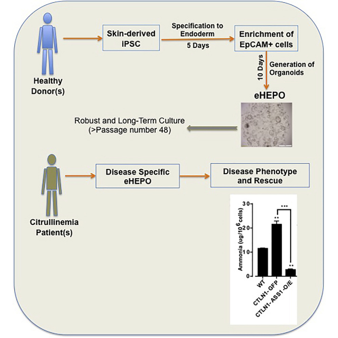
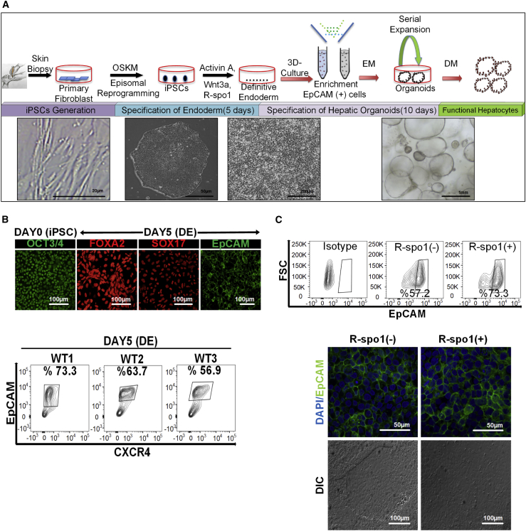
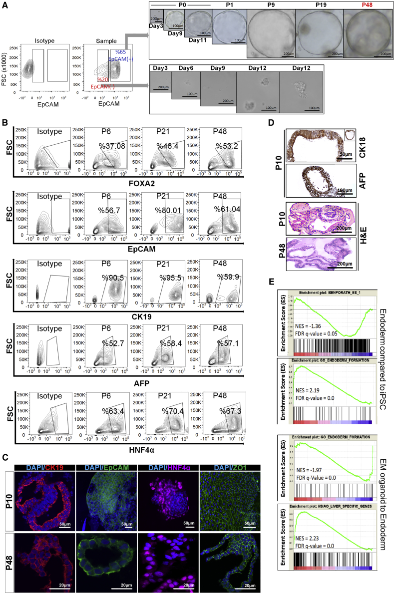
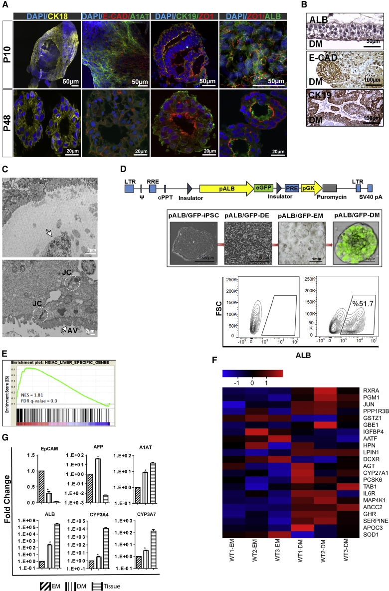
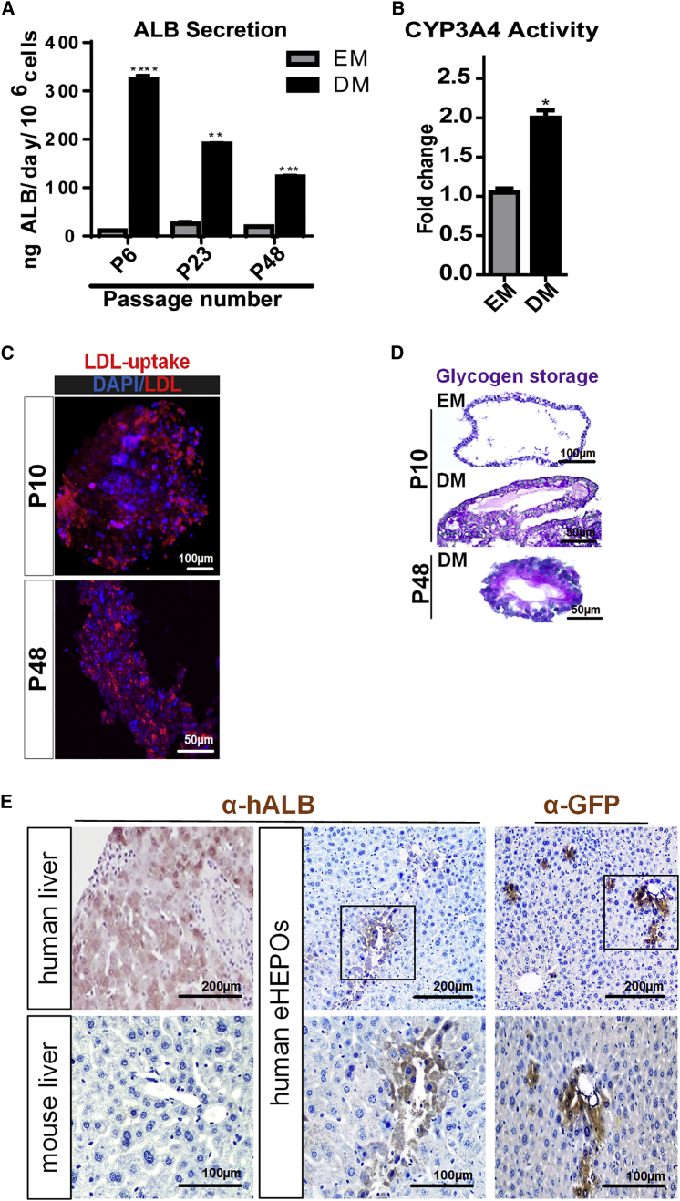
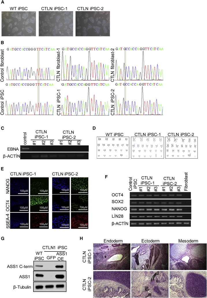
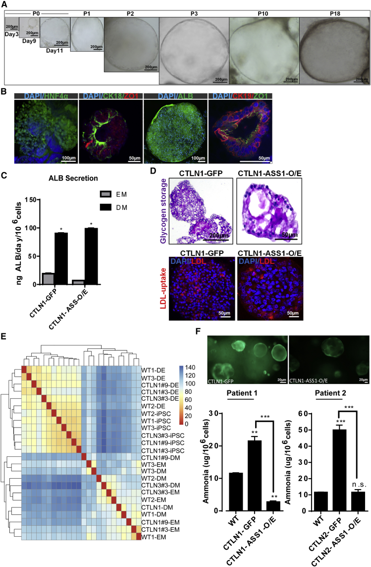
Organoid technologies have become a powerful emerging tool to model liver diseases, for drug screening, and for personalized treatments. These applications are, however, limited in their capacity to generate functional hepatocytes in a reproducible and efficient manner. Here, we generated and characterized the hepatic organoid (eHEPO) culture system using human induced pluripotent stem cell (iPSC)-derived EpCAM-positive endodermal cells as an intermediate. eHEPOs can be produced within 2 weeks and expanded long term (>16 months) without any loss of differentiation capacity to mature hepatocytes. Starting from patient-specific iPSCs, we modeled citrullinemia type 1, a urea cycle disorder caused by mutations in the argininosuccinate synthetase (ASS1) enzyme. The disease-related ammonia accumulation phenotype in eHEPOs could be reversed by the overexpression of the wild-type ASS1 gene, which also indicated that this model is amenable to genetic manipulation. Thus, eHEPOs are excellent unlimited cell sources to generate functional hepatic organoids in a fast and efficient manner.

Keywords: 3D organoid; EpCAM; citrullinemia; disease modelling; hepatocyte; hepatocyte differentiation; iPSC; liver.


Figures

None
Graphical abstract
Figure 1
Figure 1
Generation of EpCAM+ Progenitors from hiPSCs as an Intermediate of Endoderm-Derived Hepatic Organoids (A) Schematic representation and timeline of organoid culture establishment. (B) Upper panel: expression of OCT3/4 (pluripotency marker), FOXA2, SOX17, and EpCAM (endoderm markers), in the different stages by immunostaining (20× magnification). Lower panel: flow-cytometric analysis of EpCAM and CXCR4 to assess the efficiency of endoderm generation at day 5. Three independent healthy iPSC lines (wild-type 1 [WT1], WT2, WT3) were tested for endoderm induction based on EpCAM and CXCR4 positivity (n = 3 for each experiment). (C) Upper panel: representative flow-cytometric analysis with isotype control demonstrating the increase in EpCAM-expressing cell number with addition of R-spo1. Experiments were performed in triplicates for three different iPSC lines derived from healthy donors. Lower panel: effect of R-spo1 on the morphology of iPSC derived endodermal cells on 2D culture. DIC, differential interference contrast.
Figure 2
Figure 2
Establishment of Endoderm-Derived Hepatic Organoids (A) Organoid formation potency of sorted EpCAM+ and EpCAM cells and bright-field images of organoid culture from various passages, where P indicates passage number. (B) Phenotypic characterization of endoderm-derived WT organoid culture at different passage numbers (p6, p21, and p48) during expansion on EM. (C) Confocal images of organoids for EpCAM, CK19, and HNF4α. Nuclei were co-stained with DAPI. In the dataset for p10 organoids, EpCAM∖HNF4α and ZO-1 were stained in the whole-mount organoids. The others were stained from frozen sections. (D) Immunohistochemical staining of CK18 and AFP in organoids. H&E indicates hematoxylin-eosin staining. (E) GSEA plots for differentially expressed genes during iPSC to endoderm induction and endoderm to organoid differentiation. NES and FDR q values are listed for each gene set analyzed.
Figure 3
Figure 3
In Vitro Differentiation of Endoderm-Derived Hepatic Organoids into Mature Hepatocytes (A) Confocal images of organoids cultured for 10–14 days in DM conditions stained for CK18, E-cadherin, A1AT, ZO1, and ALB. Nuclei were stained with DAPI. In the dataset for p10 organoids, CK18, E-CAD/A1AT, and ZO-1/ALB were stained in the whole-mount organoids. The others were stained from frozen sections. (B) Immunohistochemical staining of organoids for ALB, CK19, and E-cadherin. (C) Transmission electron microscopy image of an endoderm-derived hepatic organoid (eHEPO). Arrow shows apoptotic and multivesicular bodies (upper panel). White circles and arrow indicate intercellular junctional complexes (JC) and apical villus (AV), respectively (lower panel). (D) Lentiviral albumin promoter-GFP reporter to monitor albumin expression in organoids. Representative bright-field and fluorescence microscopy images of pALB-GFP reporter bearing iPSCs at the indicated stages of differentiation. Flow-cytometry analysis to quantify ALB+ cells within organoid. (E) GSEA analysis of differentially regulated genes in DM versus EM conditions. NES and FDR q value are listed for the liver-specific gene set analyzed. (F) Heatmap showing the expression of EM- and DM-related genes. (G) qPCR-based mRNA expression analysis of indicated genes in EM and DM organoids as well as human liver tissue. Fold changes were calculated as DM/EM and/or tissue/EM (n = 4 for each of three separate differentiation) (p ≤ 0.05).
Figure 4
Figure 4
In Vitro and In Vivo Functionality of Endoderm-Derived Hepatic Organoids (A) Albumin secretion of the WT organoids from different passage numbers (p6, p23, p48) cultured in EM and DM conditions as measured by ELISA. Data are shown as mean ± SD of n = 3, given as ngALB/day/million cells. (B) CYP3A4 activity in organoids cultured in EM and DM conditions expressed as RLU/mL/million cells. (C) Uptake of low-density lipoprotein (LDL) detected on day 14 by immunofluorescence staining in WT DM organoids from p10 and p48. (D) Glycogen storage function of WT DM organoids from p10 and p48 by periodic-acid-Schiff (PAS) staining (magenta). EM condition was used as undifferentiated control. (E) Immunohistochemistry with anti-GFP and anti-human albumin antibodies in DMN-treated NSG mouse liver sections transplanted with mature eHEPOs. The presence of GFP+ and human ALB+ cells demonstrates engraftment of hepatocytes into mouse liver. Error bars in (A) and (B) denote ±SD of three independent experiments (p ≤ 0.05; ∗∗p ≤ 0.01; ∗∗∗p ≤ 0.001).
Figure 5
Figure 5
Derivation and Characterization of Citrullinemia-Specific hiPSCs (A) Morphology of WT iPSC, CTLN iPSC-1, and CTLN iPSC-2. (B) Sequence analysis of ASS1 exon15 mutations in healthy and patient-derived fibroblasts and the iPSCs. (C) PCR-based integration analysis of the episomal reprogramming vectors. (D) Karyotype analysis of WT iPSC, CTLN iPSC-1, and CTLN iPSC-2. (E) Immunofluorescence images of iPSCs stained for NANOG, OCT4, and SSEA-4. Cell nuclei were counterstained with Hoechst. Scale bars, 100 μm. (F) mRNA expression levels of pluripotency-related genes. (G) Western blot for ASS1 using two independent antibodies in WT iPSCs, CTLN1 iPSC-overexpressing GFP, or ASS1 cDNA (OE). (H) Teratoma formation of CTLN iPSC-1 and CTLN iPSC-2 in SCID mice. Scale bars, 100 μm.
Figure 6
Figure 6
Endoderm-Derived Hepatic Organoids for Modeling Citrullinemia (A) Bright-field image of citrullinemia patient-derived hepatic organoid cultures. (B) Confocal images of CTLN organoids stained for HNF4α, CK18, ZO-1, CK19, and ALB. Nuclei were stained with DAPI. In the dataset for CTLN organoids, HNF4α and ALB were stained in the whole-mount organoids. The others were stained from frozen sections. (C) In vitro functionality of CTLN-GFP and CTLN-ASS-O/E organoids. Albumin secretion of CTLN1-GFP and CTLN1-ASS-O/E organoids at p10 cultured in EM and DM conditions measured by ELISA. Error bars denote ±SD of three independent experiments. (D) Glycogen storage function of CTLN1-GFP and CTLN1-ASS-O/E organoids at p10 in DM condition by PAS staining. LDL uptake of CTLN1-GFP and CTLN1-ASS-O/E organoids at p10 in DM condition. (E) Cluster heatmap of WT and CTLN-iPSCs endoderm and organoids. (F) Ammonia detoxification capacity of healthy donor (WT indicates organoids with wild-type ASS1 gene) and patient-derived mature eHEPOs expressing either GFP (control) or ASS1-O/E as determined by ammonia elimination assay. Data are shown as ±SD of three independent experiments in two different patient-derived eHEPOs given as μg/day/million cells (p ≤ 0.05; ∗∗p ≤ 0.01; ∗∗∗p ≤ 0.001).

References

    1. Barker N., Huch M., Kujala P., van de Wetering M., Snippert H.J., van Es J.H., Sato T., Stange D.E., Begthel H., van den Born M. Lgr5 +ve stem cells drive self-renewal in the stomach and build long-lived gastric units in vitro. Cell Stem Cell. 2010;6:25–36. - PubMed
    1. Basak O., Beumer J., Wiebrands K., Seno H., van Oudenaarden A., Clevers H. Induced quiescence of Lgr5+ stem cells in intestinal organoids enables differentiation of hormone-producing enteroendocrine cells. Cell Stem Cell. 2017;20:177–190.e4. - PubMed
    1. Baxter M., Withey S., Harrison S., Segeritz C.-P., Zhang F., Atkinson-Dell R., Rowe C., Gerrard D.T., Sison-Young R., Jenkins R. Phenotypic and functional analyses show stem cell-derived hepatocyte-like cells better mimic fetal rather than adult hepatocytes. J. Hepatol. 2015;62:581–589. - PMC - PubMed
    1. Broutier L., Andersson-Rolf A., Hindley C.J., Boj S.F., Clevers H., Koo B.K., Huch M. Culture and establishment of self-renewing human and mouse adult liver and pancreas 3D organoids and their genetic manipulation. Nat. Protoc. 2016;11:1724–1743. - PubMed
    1. Carmon K.S., Gong X., Lin Q., Thomas A., Liu Q. R-spondins function as ligands of the orphan receptors LGR4 and LGR5 to regulate Wnt/β-catenin signaling. Proc. Natl. Acad. Sci. U S A. 2011;108:11452–11457. - PMC - PubMed

Publication types