. 2019 Nov 5;30(5):890-902.e8.
doi: 10.1016/j.cmet.2019.08.012. Epub 2019 Sep 12.

Mediation of the Acute Stress Response by the Skeleton

Affiliations

Mediation of the Acute Stress Response by the Skeleton

Julian Meyer Berger et al. Cell Metab. .

Abstract

We hypothesized that bone evolved, in part, to enhance the ability of bony vertebrates to escape danger in the wild. In support of this notion, we show here that a bone-derived signal is necessary to develop an acute stress response (ASR). Indeed, exposure to various types of stressors in mice, rats (rodents), and humans leads to a rapid and selective surge of circulating bioactive osteocalcin because stressors favor the uptake by osteoblasts of glutamate, which prevents inactivation of osteocalcin prior to its secretion. Osteocalcin permits manifestations of the ASR to unfold by signaling in post-synaptic parasympathetic neurons to inhibit their activity, thereby leaving the sympathetic tone unopposed. Like wild-type animals, adrenalectomized rodents and adrenal-insufficient patients can develop an ASR, and genetic studies suggest that this is due to their high circulating osteocalcin levels. We propose that osteocalcin defines a bony-vertebrate-specific endocrine mediation of the ASR.

Keywords: Glast; Vglut2; adrenal; bone; fight or flight; glutamate; osteoblast; osteocalcin; parasympathetic; stress response.


Conflict of interest statement

Declaration of Interests

The authors declare no competing financial interests.

Figures

Figure 1.
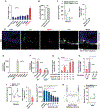
Figure 1.. Stressors trigger a rapid surge of circulating bioactive osteocalcin (Ocn) in rodents and humans.
(A) Serum Ocn levels in 3-month-old WT mice after restraint or foot shocks. (B-E) Serum Ocn levels in 2- and 6-month-old, male and female WT mice, at 12 p.m. and 8 p.m., and C57Bl/6 or 129SV WT mice after foot shocks. (F) Serum Ocn levels in WT female rats after restraint. (G) Serum Ocn levels in WT mice after foot shock and humans after public speaking stress. (H) Serum Ocn levels in WT mice after TMT or rabbit urine exposure. (I) Serum Ocn and corticosterone levels in TMT-exposed WT mice (gray). (J) Serum Ocn levels and rate of Ocn release in TMT-exposed WT mice expressing hM4Di in the BLA after injection of CNO or vehicle. Mice are 3-month-old females unless otherwise specified. Values are mean ±SEM. ns, not significant; *, p<.05; **, p<.01; by Student’s t-test or one-way ANOVA with Bonferroni post hoc test.
Figure 2.
Figure 2.. Bioactive osteocalcin is released from cells of the osteoblast lineage during an acute stress response.
(A) Serum Ocn levels and rate of Ocn release in nadolol- and vehicle-treated WT mice exposed to TMT. (B) Serum Ocn levels and rate of Ocn release in adrenalectomized (ADX) and sham-operated TMT-exposed WT mice. (C) Northern blot analysis of Ocn (top) and L19 (bottom) expression after foot shock. (D) Serum Ocn levels and rate of Ocn release in oc/oc and WT mice before and after TMT exposure. (E) Serum Ocn levels and rate of Ocn release in Tphvil−/− and Tphf/f mice before and after TMT exposure. (F) Serum Ocn levels and rate of Ocn release in α1(I) CollagenDTA/+ and μ1(I) Collagen+/+ mice before and after TMT exposure. (G) Serum Ocn levels and rate of Ocn release in chlorisondamine- and vehicle-treated WT mice before and after TMT exposure. Mice are 3-month-old females. Rats are 4-month-old females. Values are mean ±SEM. ns, not significant; *, p<.05; **, p<.01; by Student’s t- test or one-way ANOVA with Bonferroni post hoc test.
Figure 3.
Figure 3.. Glutamate mediates the stressor-induced release of bioactive osteocalcin from osteoblasts.
(A) Supernatant Ocn levels after 1-hour treatment of osteoblasts with indicated neurotransmitters. (B) Serum glutamate levels before and after TMT exposure. (C) Serum Ocn levels and rate of Ocn release before and after TMT stress in pegylated diphtheria toxin (PEG-DT)- or vehicle-injected Vglut2iDTR mice. (D) Immunofluorescence of femoral metaphysis of WT mice, Ob: osteoblast, N: glutamatergic neurite (scale: 5 μm). (E) Expression of glutamate transporters in osteoblasts. (F) Radiolabeled glutamate transport into Glast−/− and WT osteoblasts treated with vehicle or UCPH102. (G) Ocn levels in supernatants after 1-hour treatment with glutamate and UCPH102 of untreated or warfarin pre-treated mouse osteoblast cultures. (H) Supernatant Ocn levels 1 hour after treatment of Glast−/− and WT osteoblasts with glutamate. (I) Serum Ocn levels and rate of Ocn release in Glast−/− and WT mice before and after TMT exposure. (J) GGCX activity on Ocn in WT osteoblast lysates treated with glutamate. (K) Serum Ocn in WT mice expressing hM3Dq in the BLA after injection of CNO or vehicle. Mice are 3-month-old females. Values are mean ±SEM. nd, not detectible. ns, not significant; *, p<.05; **, p<.01; by Student’s t-test or one-way ANOVA with bonferroni post hoc test.
Figure 4.
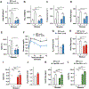
Figure 4.. Osteocalcin signaling in peripheral organs is necessary to mount an ASR.
(A-F) Energy expenditure, oxygen consumption, temperature, heart rate, arterial oxygen saturation and blood glucose in Ocn−/− and WT mice before and after foot shock. (G) Newtonian airway resistance in Ocn−/− and WT mice. (H-I) Heart rate and arterial oxygen saturation in Gprc6a−/− and WT mice before and after foot shock. (J) Newtonian airway resistance in Gprc6a−/− and WT mice. (K-L) Heart rate and arterial oxygen saturation in Gpr158−/− and WT mice before and after foot shock. Mice are 3-month-old females. Rats are 4-month-old females. Values are mean ±SEM. ns, not significant; *, p<.05; **, p<.01; by Student’s t-test or one-way ANOVA with bonferroni post hoc test.
Figure 5.
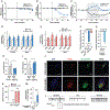
Figure 5.. Osteocalcin inhibits parasympathetic tone during an ASR.
(A) Sympathetic nerve activity in WT mice injected with Ocn (30 ng/g) or vehicle. (B) Parasympathetic nerve activity in WT mice injected with Ocn (30 ng/g) or vehicle. (C) Parasympathetic nerve activity after TMT exposure in Ocn−/− and WT littermates. (D-E) Chat, Cht1 and Vachtl expression in trachea and heart of Ocn−/−, Gprc6a−/− and WT littermates. (F) Contraction of electrically stimulated Gprc6a−/− or WT mouse tracheal rings treated with Ocn (10 ng/ml) or vehicle. (G) Contraction of electrically stimulated human tracheal rings treated with Ocn (10 ng/ml) or vehicle. (H-I) Newtonian airway resistance and heart rate in vagotomized or sham-operated Ocn−/− and WT mice. (J) Immunofluorescence of airway (scale bar: 15 μm) and cardiac ganglia of Gprc6a-Gfp mice, PG: parasympathetic ganglia (scale bar: 12 μm). (K-L) Firing frequency in WT and Gprc6a−/− tracheal post-ganglionic parasympathetic neurons before, during and after treatment with Ocn (15 ng/ml) normalized to control and representative traces of action currents in Ocn−/− or vehicle-treated WT tracheal post-ganglionic parasympathetic neurons. (M) Firing frequency in tracheal post-ganglionic parasympathetic neurons after treatment with sera from stressed Ocn−/− or WT mice. Mice are 3-month-old females. Values are mean ±SEM. ns, not significant; *, p<.05; **, p<.01; by Student’s t-test or one-way ANOVA with bonferroni post hoc test.
Figure 6.
Figure 6.. High circulating osteocalcin levels account for the ability of adrenalectomized mice to develop an ASR.
(A-E) Energy expenditure, oxygen consumption, temperature, heart rate and arterial oxygen saturation in ADX and sham-operated mice before and after foot shock. (F-G) Temperature and heart rate in ADX and sham-operated rats before and after restraint. (H) Serum Ocn levels in WT sham-operated, ADX, Ocn+/− ADX, Ocn+/− sham-operated and Ocn−/− ADX mice. (I-J) Energy expenditure and oxygen consumption in WT ADX, Ocn+/− ADX and Ocn−/− ADX mice before and after foot shock. (K-L) Heart rate and arterial oxygen saturation in WT ADX, Ocn+/− ADX and Ocn−/− ADX mice before and after foot shock. (M-O) Energy expenditure, oxygen consumption and heart rate in WT mice injected with Ocn (30 ng/g) or vehicle. (P) Schematic representation of the endocrine mediation of the ASR in bony vertebrates. Stress signaling in the amygdala results in glutamate release in bone. Glutamate enters osteoblasts through Glast, exerts a competitive inhibition on GGCX and as a result bioactive Ocn is released. Ocn binds to Gprc6a on peripheral parasympathetic neurons to inhibit their activity, allowing the ASR to begin (AP, action potential). Mice are 3-month-old females. Rats are 4-month-old females. Values are mean ±SEM. ns, not significant; *, p<.05; **, p<.01; by Student’s t-test or one-way ANOVA with bonferroni post hoc test.

Comment in

Similar articles

Cited by

References

    1. Arteaga-Solis E, Zee T, Emala CW, Vinson C, Wess J, and Karsenty G (2013). Inhibition of leptin regulation of parasympathetic signaling as a cause of extreme body weight-associated asthma. Cell Metab 17, 35–48. - PMC - PubMed
    1. Axelrod J, and Reisine TD (1984). Stress hormones: their interaction and regulation. Science 224, 452–459. - PubMed
    1. Azuma Y, Sato H, Oue Y, Okabe K, Ohta T, Tsuchimoto M, and Kiyoki M (1995). Alendronate distributed on bone surfaces inhibits osteoclastic bone resorption in vitro and in experimental hypercalcemia models. Bone 16, 235–245. - PubMed
    1. Brumovsky PR, Robinson DR, La JH, Seroogy KB, Lundgren KH, Albers KM, Kiyatkin ME, Seal RP, Edwards RH, Watanabe M, et al. (2011a). Expression of vesicular glutamate transporters type 1 and 2 in sensory and autonomic neurons innervating the mouse colorectum. J Comp Neurol 519, 3346–3366. - PMC - PubMed
    1. Brumovsky PR, Seroogy KB, Lundgren KH, Watanabe M, Hokfelt T, and Gebhart GF (2011b). Some lumbar sympathetic neurons develop a glutamatergic phenotype after peripheral axotomy with a note on VGLUT(2)-positive perineuronal baskets. Exp Neurol 230, 258–272. - PMC - PubMed

Publication types

MeSH terms