

Retinal inner nuclear layer volume reflects inflammatory disease activity in multiple sclerosis; a longitudinal OCT study
- PMID: 31523449
- PMCID: PMC6728683
- DOI: 10.1177/2055217319871582
Retinal inner nuclear layer volume reflects inflammatory disease activity in multiple sclerosis; a longitudinal OCT study
Abstract
Background: The association of peripapillary retinal nerve fibre layer (pRNFL) and ganglion cell-inner plexiform layer (GCIPL) thickness with neurodegeneration in multiple sclerosis (MS) is well established. The relationship of the adjoining inner nuclear layer (INL) with inflammatory disease activity is less well understood.
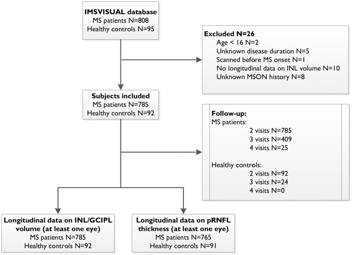
Objective: The objective of this paper is to investigate the relationship of INL volume changes with inflammatory disease activity in MS.Methods In this longitudinal, multi-centre study, optical coherence tomography (OCT) and clinical data (disability status, relapses and MS optic neuritis (MSON)) were collected in 785 patients with MS (68.3% female) and 92 healthy controls (63.4% female) from 11 MS centres between 2010 and 2017 and pooled retrospectively. Data on pRNFL, GCIPL and INL were obtained at each centre.
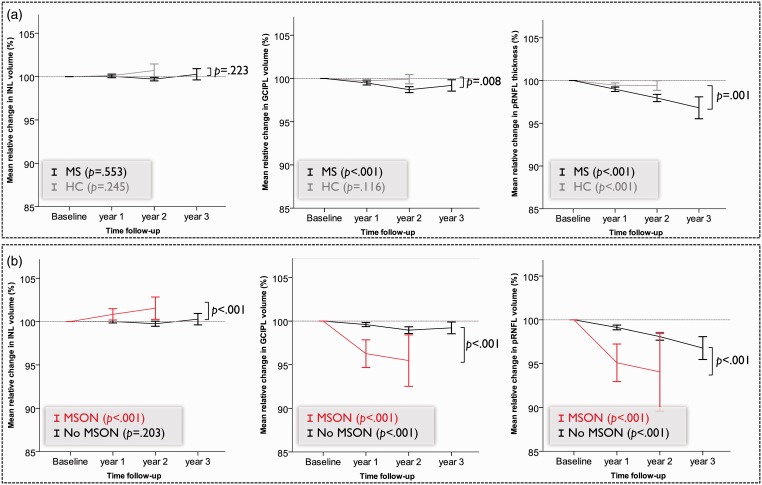
Results: There was a significant increase in INL volume in eyes with new MSON during the study (N = 61/1562, β = 0.01 mm3, p < .001). Clinical relapses (other than MSON) were significantly associated with increased INL volume (β = 0.005, p = .025). INL volume was independent of disease progression (β = 0.002 mm3, p = .474).
Conclusion: Our data demonstrate that an increase in INL volume is associated with MSON and the occurrence of clinical relapses. Therefore, INL volume changes may be useful as an outcome marker for inflammatory disease activity in MSON and MS treatment trials.
Keywords: Inflammation; inner nuclear layer; multiple sclerosis; optical coherence tomography.
Figures
References
-
- Petzold A, Balcer LJ, Calabresi PA, et al. Retinal layer segmentation in multiple sclerosis: A systematic review and meta-analysis. Lancet Neurol 2017; 16: 797–812. - PubMed
-
- Gordon-Lipkin E, Chodkowski B, Reich DS, et al. Retinal nerve fiber layer is associated with brain atrophy in multiple sclerosis. Neurology 2007; 69: 1603–1609. - PubMed
-
- Sepulcre J, Murie-Fernandez M, Salinas-Alaman A, et al. Diagnostic accuracy of retinal abnormalities in predicting disease activity in MS. Neurology 2007; 68: 1488–1494. - PubMed
LinkOut - more resources
Full Text Sources
