. 2019 Oct 16;141(41):16374-16381.
doi: 10.1021/jacs.9b07578. Epub 2019 Sep 26.

Versatile Peptide Macrocyclization with Diels-Alder Cycloadditions

Versatile Peptide Macrocyclization with Diels-Alder Cycloadditions

Jeffrey E Montgomery et al. J Am Chem Soc. .

Abstract

Macrocyclization can improve bioactive peptide ligands through preorganization of molecular topology, leading to improvement of pharmacologic properties like binding affinity, cell permeability, and metabolic stability. Here we demonstrate that Diels-Alder [4 + 2] cycloadditions can be harnessed for peptide macrocyclization and stabilization within a range of peptide scaffolds and chemical environments. Diels-Alder cyclization of diverse diene-dienophile reactive pairs proceeds rapidly, in high yield and with tunable stereochemical preferences on solid-phase or in aqueous solution. This reaction can be applied alone or in concert with other stabilization chemistries, such as ring-closing olefin metathesis, to stabilize loop, turn, and α-helical secondary structural motifs. NMR and molecular dynamics studies of model loop peptides confirmed preferential formation of endo cycloadduct stereochemistry, imparting significant structural rigidity to the peptide backbone that resulted in augmented protease resistance and increased biological activity of a Diels-Alder cyclized (DAC) RGD peptide. Separately, we demonstrated the stabilization of DAC α-helical peptides derived from the ERα-binding protein SRC2. We solved a 2.25 Å cocrystal structure of one DAC helical peptide bound to ERα, which unequivocally corroborated endo stereochemistry of the resulting Diels-Alder adduct, and confirmed that the unique architecture of stabilizing motifs formed with this chemistry can directly contribute to target binding. These data establish Diels-Alder cyclization as a versatile approach to stabilize diverse protein structural motifs under a range of chemical environments.


Conflict of interest statement

Notes

The authors declare no competing financial interests.

Figures

Figure 1.
Figure 1.
Synthesis, stability, and activity of Diels-Alder cyclized peptides. (A) Schematic of a DAC peptide functionalization. (B) Absorbance (215 nm) and extracted ion chromatogram (EIC) LC-MS traces of crude peptide following the indicated functionalization and reaction times with the RGD compound 1 scaffold. (C) Representative reaction time course quantifying conversion of 1 to cyclized 1a/1b isomers. (D) Extended time course tracking of HPLC-purified 1a incubated with βME in aqueous buffer, verifying cycloadduct stability (βME-trapped species m/z = 914.3). (E) In vitro trypsin and chymotrypsin resistance assays for 1a versus linear analog 1wt, and 4a and linear analog 4wt, respectively. (F) Cell migration model scratch wound assay comparing reduction in wound closure by RGD peptides 1wt and 1a at 10 and 20 μM relative to DMSO control. Data represent mean ± s.d. distance measurements from images of 2 biological replicates, 5 distance measurements each. n.s. = not significant; * = P < 0.05; ** = P < 0.0005; Student’s t-test.
Figure 2.
Figure 2.
NMR spectroscopy characterization and MD simulations of DAC peptide 4a and linear precursors. (A) 1H-NMR spectra of 4wt (black), 4hex (blue), and 4a (red) in the vinylic (~6–5 p.p.m.) region. (B) Model of 4a cycloadduct depicting through-bond and through-space interactions between cycloadduct protons as determined by TOCSY and NOESY (orange: bridgehead interactions; purple: methyl-group interactions; black: Cys-side chain interactions; red: vinylic interactions). (C) 3JH-N-Cα-Hα coupling constant values for each residue of the three peptides studied. (D) Peptide backbone alignment over 50 ns simulations for 4wt and 4a. (E) RMSD values for 4wt and 4a peptide backbones and all atoms (excluding hydrogens).
Figure 3.
Figure 3.
Synthesis, stability, and secondary structural characterization of Diels-Alder cyclized p53 peptide. (A) Schematic of DAC-p53 peptide synthesis. (B) HPLC trace and resulting aqueous solution reaction time course of conversion of 5 to cyclized isomers 5a-c. (C) Incubation of DAC-p53 peptide 5 mixture with βME in aqueous buffer, verifying Michael adduction of linear precursor 5 and stability of cyclized isomers. (D) CD spectra of linear analog 5wt and 5a-c.
Figure 4.
Figure 4.
Synthesis, secondary structural characterization and activity of Diels-Alder cyclized and double-stapled SRC2 peptides. (A) Sequence of SRC2 wildtype peptide used as scaffold with conserved LXXLL motif; staple placement and structures of DAC-SRC2 peptides 6a-b and 7a and double-stapled 8a. (B) HPLC traces of pre-cursor peptide used and resulting cyclized product profile of compound 7 following 4 hr on-resin heating in DMSO. (C) CD spectra of isolated DAC-SRC2 peptides compared to unmodified wildtype sequence. (D) Synthetic scheme for layering ring-closing metathesis and Diels-Alder cyclization for the synthesis of double-stapled SRC2 peptide 8a. (E) HPLC traces of pre-cursor peptide 8pre, ring-closing metathesis product 8rcm, Diels-Alder functionalized peptide 8 and fully-DAC-SRC2 double-stapled peptide 8a. (F) CD spectra of helical compound 8a. (G) Normalized FP assay results showing a dose range of DAC-SRC2 peptides effectively competing off fluorescein-labeled wildtype SRC peptide from wildtype ERα. Each data point represents measurements from two independent experiments run in triplicate, ±s.e.m. Dose-response curves fit with a four parameter log(inhibitor) vs response equation in Prism 5 graphing software.
Figure 5.
Figure 5.
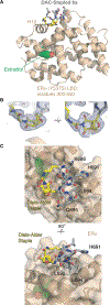
X-ray crystal structure of Diels-Alder cyclized SRC2 peptide 6a bound to estrogen receptor-α (PDB: 6PIT). (A) LBD of ERα Y537S mutant protein bound with estradiol and 6a located in the AF2 cleft. (B) Two views of the structure of the DAC-SRC2 peptide 6a cycloadduct (C = yellow, N = blue, O = red) with endo stereochemistry overlaid with electron density map (grey grid) from resolved crystallographic data (2mFo-DFc map contoured to 1σ). (C) Dorsal (top) and axial (bottom) view of 6a bound in the AF2 binding pocket. Relevant residues numbered according to SRC2 sequence are highlighted, as well as the Diels-Alder cyclization moiety in yellow. Flexible portions of the structure with low electron density are omitted.

References

    1. Bullock BN; Jochim AL; Arora PS, Assessing Helical Protein Interfaces for Inhibitor Design. Journal of the American Chemical Society 2011, 133 (36), 14220–14223. - PMC - PubMed
    1. Gavenonis J; Sheneman BA; Siegert TR; Eshelman MR; Kritzer JA, Comprehensive analysis of loops at protein-protein interfaces for macrocycle design. Nature Chemical Biology 2014, 10, 716. - PMC - PubMed
    1. Spokoyny AM; Zou Y; Ling JJ; Yu H; Lin Y-S; Pentelute BL, A Perfluoroaryl-Cysteine SNAr Chemistry Approach to Unprotected Peptide Stapling. Journal of the American Chemical Society 2013, 135 (16), 5946–5949. - PMC - PubMed
    1. Guillen Schlippe YV; Hartman MCT; Josephson K; Szostak JW, In Vitro Selection of Highly Modified Cyclic Peptides That Act as Tight Binding Inhibitors. Journal of the American Chemical Society 2012, 134 (25), 10469–10477. - PMC - PubMed
    1. Hill TA; Shepherd NE; Diness F; Fairlie DP, Constraining Cyclic Peptides To Mimic Protein Structure Motifs. Angewandte Chemie International Edition 2014, 53 (48), 13020–13041. - PubMed

Publication types